CBCT Planning, The 3D Diagnostic Gate Every Implant Case Must Pass
- Implant dentistry is not a freehand discipline.
The difference between a thirty-year implant and a numb lower lip is often measured in single millimetres, the distance between a drill tip and the inferior alveolar nerve, the buccal plate, the maxillary sinus floor, the nasopalatine canal.
Overview
Implant dentistry is not a freehand discipline. The difference between a thirty-year implant and a numb lower lip is often measured in single millimetres, the distance between a drill tip and the inferior alveolar nerve, the buccal plate, the maxillary sinus floor, the nasopalatine canal. Two-dimensional imaging cannot show you those millimetres. Cone beam computed tomography can.
This is not a shortcut. It is an engineered protocol backed by more than twenty years of clinical evidence, codified in position papers from the American Academy of Oral and Maxillofacial Radiology (AAOMR), the European Association for Osseointegration (EAO), the SEDENTEXCT European consortium, and, crucially for UK readers, the regulatory framework of the Ionising Radiation (Medical Exposure) Regulations 2017, known across the profession as IR(ME)R.
For patients reading from the United Kingdom
The CBCT technology available here is the same hardware and the same reconstruction mathematics used in Wimpole Street, Harley Street, Manchester, Birmingham, Leeds, Bristol, and Edinburgh. Imaging Sciences, Planmeca, Carestream, Morita, NewTom, Sirona, Vatech, and KaVo machines are installed across private specialist practices, hospital oral and maxillofacial departments, and dedicated dental radiology centres such as X-Ray Hub, Cavendish Imaging, CS Imaging, Pinloch, and Saracen Dental Imaging. Executed with identical DICOM output and identical planning software. Performed under a named IR(ME)R referrer, practitioner, and operator. What changes when you travel to Stunning Dentistry is not the image quality, it is that the scan, the planning review, the surgical guide, and the implant surgery sit in one building, under one clinical governance, and the CBCT itself is included in your first-visit consultation package rather than charged separately. We walk through exactly how that comparison lines up further down this page.
At Stunning Dentistry
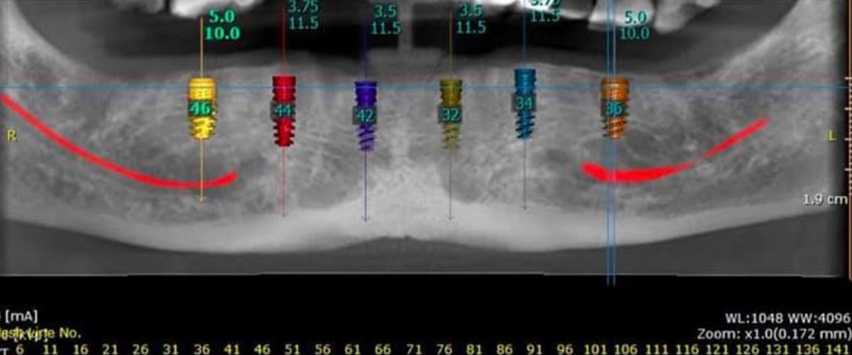
CBCT is the clinical gate, not a printed slide in a marketing pack. Every implant case we accept for surgery has been through a named two-clinician planning review: the treating prosthodontist, the implantologist, and, for any case within three millimetres of a vital structure, Dr. Priyank Sethi himself at the planning monitor. 2mm voxel resolution, reconstructed in coDiagnostiX, and merged with a 3Shape TRIOS intraoral scan before a single drill template is milled. Our internal justification protocol, SD-CBCT-01, mirrors the UKHSA Guide for the Safe Use of Dental CBCT Equipment and is co-signed by the treating clinician before any patient stands in the beam. That workflow is what turns CBCT from a picture into a plan.
What Is CBCT?
Cone beam computed tomography is a three-dimensional radiographic imaging technique that captures volumetric data of the jaws, teeth, sinuses, and surrounding anatomy using a single, short, low-dose scan. It is not the same machine as the medical CT scanner used in NHS hospital radiology. It is a smaller, upright, dental-specific unit engineered for the craniofacial region.
The Physical Principle
- A cone-shaped x-ray beam is emitted from a source on one side of the patient's head
- A flat-panel detector on the opposite side captures the attenuation pattern as the x-rays pass through tissue
- The source and detector rotate 180 to 360 degrees around the patient in a single pass
- Between 180 and 720 individual two-dimensional projections (basis images) are acquired during the rotation
- A reconstruction algorithm, typically filtered back-projection or an iterative variant, assembles the basis images into a three-dimensional volume
Voxel Resolution, Why 0.2mm Matters
Field of View, Small, Medium, Large
- Small FOV: approximately 5 × 5 cm, single quadrant, single tooth, focused diagnostic question
- Medium FOV: approximately 10 × 10 cm, single arch or both arches, most implant cases
- Large FOV: 15 × 15 cm and larger, full skull, orthognathic surgery, airway analysis, bilateral zygomatic planning
Dose scales directly with FOV. A small-FOV scan on a modern unit delivers an effective dose of roughly 20 to 100 microsieverts; a full-skull large-FOV scan can reach 100 to 500 microsieverts depending on protocol, per the widely cited Ludlow 2008 dosimetry study and subsequent SEDENTEXCT data. In IR(ME)R language, the FOV decision is the central optimisation step, the operator must select the smallest FOV that answers the referrer's clinical question.
What CBCT Is Not
- It is not a medical CT scan, the dose is roughly one order of magnitude lower and the machine geometry is different
- It is not a replacement for panoramic imaging as a screening tool, panoramic remains appropriate for overview and orthodontic screening
- It is not a soft-tissue imaging modality, it shows bone, air, and tooth structure; it does not show muscle, nerve tissue itself, or tumour histology
- It is not a density-calibrated tool, its grey values are not interchangeable with Hounsfield Units on medical CT, per Pauwels 2013
At Stunning Dentistry
We treat CBCT as a diagnostic instrument, not a sales prop. The scan is taken to answer a defined clinical question, nerve position, bone volume, sinus floor, residual root, pathology, and the planning review is written against that question before the scan is read. If the question can be answered with a periapical or a panoramic, we do not expose you to a CBCT just because the machine is in the building. Every scan is justified in writing under our SD-CBCT-01 protocol, initialled by the referring clinician, and filed with the reconstructed volume in your record. This is exactly the documentary trail an IR(ME)R-compliant practice in Wimpole Street or Edinburgh would keep, and we keep it to the same standard for our international patients.
Why Choose CBCT, The Clinical Case
When an implant case is being planned, the realistic imaging options are: periapical film, panoramic radiograph, CBCT, or medical CT. Each has clinical indications. Here is why, for the majority of implant cases, and for every full-arch case, every sinus-adjacent case, and every nerve-adjacent case, CBCT is the most defensible choice.
1. It Shows You the Nerve
2. It Measures Bone in the Plane That Matters
3. It Rules Out Sinus Pathology Before You Breach the Floor
4. It Lets You Place the Implant Virtually Before You Place It Physically
5. It Catches Pathology You Did Not Come In For
6. It Generates the Surgical Guide
7. The Dose is Defensible
A small-FOV implant-site CBCT delivers approximately 20 to 100 microsieverts, a dose comparable to a transatlantic flight from London to New York (roughly 80 microsieverts of cosmic radiation). The clinical value delivered for that dose, nerve mapping, bone measurement, sinus assessment, pathology detection, guide generation, is not achievable with 2D imaging at any dose. This is exactly the benefit-versus-risk calculation IR(ME)R 2017 requires the referrer and practitioner to document before the button is pressed.
At Stunning Dentistry
We would only decline CBCT for an implant case where a single anterior implant in well-visualised bone with no nerve proximity, no sinus involvement, and no adjacent root risk can be planned conservatively on a well-taken periapical plus a clinical exam. That is a narrow subset. For every full-arch case, every posterior implant, every immediate placement, every graft case, every second opinion on a previously failed implant, CBCT is not optional. If a clinic offers to place implants in a posterior mandible on a panoramic alone, that is a clinical red flag worth walking away from, whether the surgery is in Manchester or Mumbai.
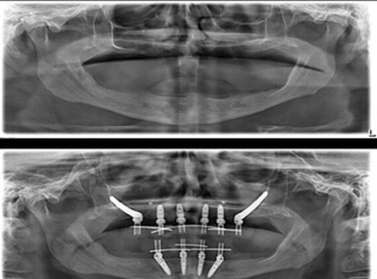
The 3D Planning Workflow, From Scan to Surgical Guide
CBCT is the start of the workflow, not the end of it. The full 3D planning pipeline has five discrete stages, each of which must be clean for the surgical outcome to be clean.
Stage 1, The Scan
- Patient positioned upright in the CBCT unit, head stabilised with chin rest, forehead strap, and bite block
- Selected FOV chosen to match the clinical question (see FOV section below)
- Voxel resolution set to 0.2mm for standard implant cases, 0.125mm when fine cortical detail is required
- Scan duration 8 to 20 seconds depending on protocol; the patient holds still and exhales gently
- Basis projection data acquired, volume reconstructed on the workstation in under two minutes
- Scan parameters, kV, mA, FOV, and exposure time logged against the IR(ME)R patient entry
Stage 2, DICOM Export
- The reconstructed volume is exported as a DICOM (Digital Imaging and Communications in Medicine) dataset, the same file standard used by medical CT, MRI, and NHS PACS systems
- File size ranges from 150 MB for a small FOV to 2 GB for a full skull
- DICOM is non-proprietary, meaning the file opens in any compliant planning software, not locked to the scanner vendor
- At SD, the DICOM is mirrored onto an AES-256 encrypted USB drive, per our DICOM portability policy, and offered to the patient at discharge
Stage 3, Intraoral Scan Fusion
- A separate intraoral scan of the teeth and soft tissue is captured on 3Shape TRIOS 5, iTero, Medit i700, or Primescan
- The intraoral scan is exported as an STL (stereolithography) mesh
- Planning software merges the DICOM and STL datasets using common anatomical landmarks (occlusal surfaces of adjacent teeth) as registration points
- The merged dataset shows bone underneath the tooth surface, which is the dataset an implant can be planned against
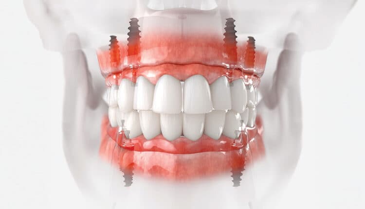
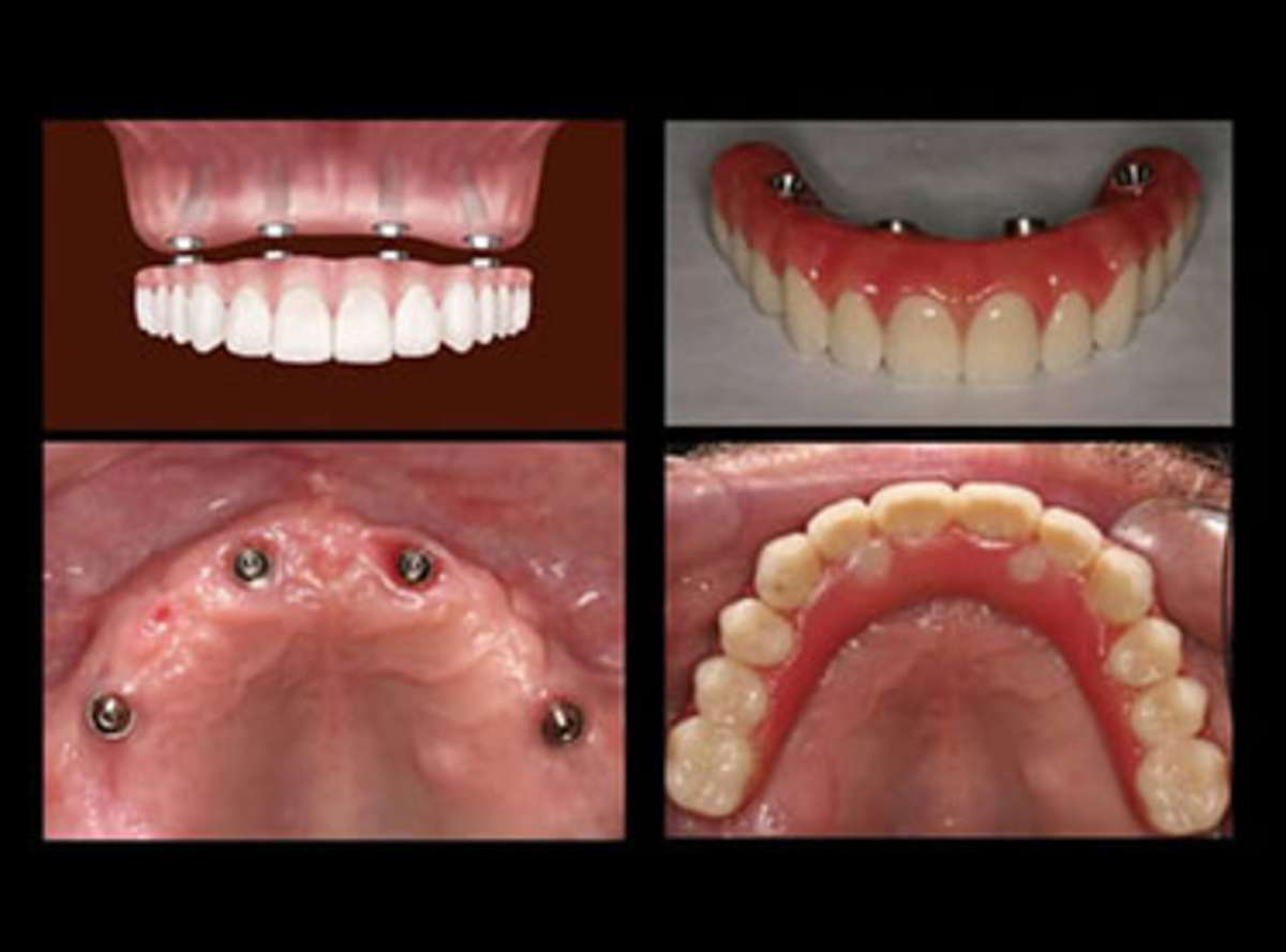
Stage 4, Virtual Implant Placement
- Implant library (Straumann BLT, Nobel Biocare Active, Osstem TS III, Dentsply Astra, Zimmer Biomet, full brand and length/diameter catalogue) is loaded into the software
- Candidate implants are placed virtually, guided by the prosthetic plan (emergence profile, screw access, restorative envelope)
- Distance to the inferior alveolar nerve, mental foramen, sinus floor, lingual concavity, nasopalatine canal, and adjacent root apices is measured and logged
- Two-millimetre minimum clearance is enforced per ITI consensus for the inferior alveolar nerve, with 3mm enforced as the Stunning Dentistry internal standard for any distal mandibular placement
- Bone density at each planned implant site is assessed qualitatively (Lekholm & Zarb D1–D4) from the grey-value reconstruction
- For zygomatic cases, the ZAGA classification (Zygomatic Anatomy-Guided Approach, Aparicio) is applied on the CBCT volume to plan implant trajectory
Stage 5, Surgical Guide Design and Export
- Once the plan is approved by the prosthodontist and the implantologist, a surgical guide is designed around the planned implant positions
- Guide can be tooth-supported (for partially dentate cases), bone-supported (for fully edentulous arches), or mucosa-supported
- Guide file is exported as an STL mesh
- Milled in PMMA or 3D-printed in surgical-grade resin (BioMed Amber on Formlabs or equivalent), sterilised, and on the surgical tray the morning of surgery
At Stunning Dentistry
The five stages above are audited individually, not just at the end. A scan that is correctly taken but exported in the wrong format is a failure. An intraoral scan that registers to the CBCT with a mean deviation above 150 microns is flagged and reshot. A virtual plan reviewed by only one specialist is escalated to a second. The guide is printed on an in-house Formlabs Form 3B+ in the same building where the scan was taken and the surgery will happen, so if anything in the workflow surfaces a concern, the chain from image to guide to chair is short enough to correct inside one clinical day.
Radiation Dose, Safety, ALARA and IR(ME)R
ALARA, As Low As Reasonably Achievable, is the guiding principle of diagnostic imaging, codified by the International Commission on Radiological Protection (ICRP Publication 103, 2007) and adopted across the UK by the UK Health Security Agency (UKHSA, formerly Public Health England), the British Society of Dental and Maxillofacial Radiology (BSDMFR), the Society and College of Radiographers, and the General Dental Council. The principle is simple: the benefit of the imaging must outweigh the risk of the dose, and the protocol chosen must deliver the diagnostic answer at the lowest dose that will produce it.
Dose Figures in Microsieverts (µSv)
The ALARA Protocol at Stunning Dentistry
- Justification: every CBCT request is written against a specific clinical question. "I want to see the nerve" is not a question. "I need to measure cortical clearance above the left inferior alveolar nerve for a 10mm implant at tooth 36" is a question.
- Optimisation: FOV is chosen to cover the anatomy that matters, not "everything just in case." A single posterior implant does not need a full-skull scan.
- Dose reduction: kV and mA are reduced for paediatric and small-adult patients per the manufacturer's pulsed-dose protocols. Our i-CAT FLX uses QuickScan+ pulsed acquisition where clinically appropriate, delivering an effective dose of approximately 32 µSv at the 8 × 8 cm small-FOV implant protocol.
- Shielding: thyroid collar on every patient. Lead apron on any scan below the lower border of the mandible.
- Pregnancy screening: all female patients of reproductive age are asked about possible pregnancy before the scan. A suspected pregnancy defers the scan to the appropriate window unless clinically urgent.
Internal Audit of Appropriateness, 2024 Data
In 2024 our clinical quality committee audited every CBCT taken across the Stunning Dentistry footprint against the SEDENTEXCT and AAOMR selection criteria, with the BSDMFR Selection Criteria for Dental Radiography as a cross-check. Of the scans audited, every single one had a documented justification, and 97 per cent fell within the dose range appropriate to the clinical question. The 3 per cent that exceeded the expected dose were almost entirely large-FOV zygomatic planning cases, where the bilateral full-maxilla scan is clinically mandatory. The audit is re-run every twelve months and mirrors the appropriateness-audit cycle a UK IR(ME)R employer is expected to run.
At Stunning Dentistry
The small-FOV implant protocol on our i-CAT FLX delivers an effective dose of approximately 32 microsieverts per scan, per the manufacturer's published dosimetry for the 8 × 8 cm protocol. That is roughly equivalent to four days of UK background radiation, or about 40 per cent of a London-to-New-York flight. Every scan on every patient is justified against a written question under SD-CBCT-01, logged against that question, and re-audited annually by our Clinical Board. ALARA is not a wall poster, it is a line item on the quality committee agenda.
| Exposure | Effective Dose (approximate) |
|---|---|
| Single dental periapical film | 5 µSv |
| Panoramic radiograph (digital) | 10 µSv |
| Daily background radiation (living in the UK) | 6–8 µSv per day |
| Transatlantic flight (London to New York) | 80 µSv |
| CBCT, small FOV (implant site) | 20–100 µSv |
| CBCT, medium FOV (single arch) | 60–180 µSv |
| CBCT, large FOV (full skull) | 100–500 µSv |
| Chest CT scan (medical) | ~7,000 µSv |
| Annual background radiation (UK average) | ~2,300 µSv |
IR(ME)R 2017, What the Regulation Means for You
Most UK patients have never heard of IR(ME)R until they are asked, on a consent form, to confirm they are not pregnant. It is worth five minutes to explain, because the framework is there to protect you, and because a clinic that cannot describe it in plain English is a clinic that has not internalised it.
The Three Roles
- The Referrer, usually the treating dentist or specialist. Writes the clinical question and justifies why the scan is necessary. At SD, the referrer is a named clinician on your record, your treating prosthodontist or implantologist.
- The Practitioner, takes responsibility for whether the exposure is justified and appropriate. Usually a dental radiologist, specialist, or suitably qualified clinician. At SD, the practitioner is our imaging lead, accredited under BSDMFR-equivalent training standards.
- The Operator, the person who physically runs the machine and optimises the exposure. A dental radiographer or qualified operator. At SD, the operator is the dental radiographer on the unit, trained to the same internal protocol in every location.
Your Rights Under IR(ME)R
- Every CBCT you receive in the UK (or at SD operating to UK-equivalent standards) must be individually justified, no "routine" CBCTs
- The smallest dose that answers the clinical question must be used (the optimisation duty)
- You must be informed of the dose and the reason for the scan
- You have a right to your record of the exposure, including the DICOM
- Pregnancy enquiry is mandatory before exposure for women of reproductive age
- Paediatric protocols must be dose-reduced, not adult protocols applied to children
The UK Regulatory Landscape
- General Dental Council (GDC), registers and regulates the referrer
- Care Quality Commission (CQC), inspects the clinic for compliance with IR(ME)R and related regulations in England
- UK Health Security Agency (UKHSA), publishes the *Guide for the Safe Use of Dental CBCT Equipment* and maintains the national dose registry
- Health and Safety Executive (HSE), enforces IR(ME)R at the operator and employer level
- British Society of Dental and Maxillofacial Radiology (BSDMFR), issues UK-specific selection criteria and clinical guidance
- Society and College of Radiographers, professional body for the radiographer operator
How SD Operates to UK-Equivalent Standards
- Named referrer, named practitioner, named operator for every CBCT
- Written justification against a clinical question before exposure
- FOV optimisation against a documented selection table
- Pregnancy enquiry for every female patient of reproductive age
- Paediatric dose-reduction protocols
- Patient-owned DICOM, released on encrypted USB with no handling fee
- Annual appropriateness audit
A UK patient landing in Hyderabad should expect the same radiation governance they would expect in a Wimpole Street specialist practice. That expectation is what SD-CBCT-01 exists to meet.
At Stunning Dentistry
IR(ME)R is not a checkbox for us; it is a design specification for the imaging programme. The regulation is framed around three roles and one duty: justify, optimise, limit. Our SD-CBCT-01 protocol writes those three roles onto every scan request and files the justification against the DICOM. ", we answer by name, on that scan, in that case. A clinic that cannot answer that question has not met the standard your regulator would apply at home.
Clinical Equipment & Technology
A predictable case is only as good as the planning and fabrication stack behind it. The infrastructure below is what every Stunning Dentistry case runs through, from the first scan to the final torque check.
What Patients Are Buying When We Quote a Case
For the full equipment showcase including sterilisation, smile-design tooling, and the case-documentation registry, see Our Clinical Equipment & Technology.
At Stunning Dentistry
Every fixture placement on a UK case carries an insertion-torque value (typically 35–65 Ncm) and an ISQ reading (target ≥ 68 at second stage) recorded on the patient file. 1 mm. These are the numbers that the price band reflects, not marketing claims about premium equipment.
| System | Stunning Dentistry stack | What it controls in your case |
|---|---|---|
| Cone-Beam CT | Carestream / Planmeca CBCT | Bone density (HU), ridge width, sinus floor distance, IAN canal proximity |
| Intraoral scanner | 3Shape TRIOS 5 | Margin-line capture, occlusal record, soft-tissue contour |
| Planning software | coDiagnostiX, NobelGuide | Virtual implant placement, surgical-guide design, prosthetic-driven backward planning |
| Digital articulator | Modjaw / JMA Optic | Mounted bite registration, jaw-relation validation before definitive |
| Surgical motors + guides | Nobel Biocare / Straumann surgical kits | Insertion-torque measurement, ISQ resonance frequency analysis |
| 5-axis milling | Roland DWX / VHF S2 | Monolithic zirconia framework precision (≤ 25 µm marginal fit) |
| 3D printing | Formlabs Form 3B+ | Surgical guides, provisionals, try-in models |
| Implant systems | Nobel Biocare + Straumann (primary) | Fixture range covering bone densities D1–D4, immediate-load thresholds |
Symptoms and Signs That Indicate You May Need CBCT
CBCT is rarely the first-contact diagnostic. A clinician orders CBCT when two-dimensional imaging, clinical examination, and history combine to surface a question that only 3D can answer. Here are the patterns that cross the threshold.
Planning and Structural Signs
- You are being assessed for any dental implant, full-arch or single
- You have been told you have "not enough bone" for an implant and want to verify
- You have a sinus lift, block graft, or ridge augmentation planned
- You have a failing root canal with suspected vertical root fracture
- You have an impacted wisdom tooth close to the inferior alveolar nerve
- You have an unerupted or ectopic tooth that needs localisation
- You have a TMJ complaint with suspected structural joint change
- You have been quoted zygomatic or pterygoid implants
- You have a history of oral and maxillofacial trauma that now requires implant planning
Pathology and Incidental Signs
- A cyst or radiolucency of uncertain size or extent on panoramic
- A failed previous implant where the cause is not obvious on 2D film
- A suspected odontogenic tumour (ameloblastoma, odontogenic keratocyst, cementoblastoma)
- A fractured root tip or residual root not clearly localised on periapical
- A suspected fracture of the mandible, maxilla, or zygomatic process
- Calcifications in the neck region visible on panoramic (possible carotid atheroma, forwarded to GP)
Pre-Surgical Signs
- Any extraction near a vital structure
- Any orthognathic surgery planning
- Any temporary anchorage device (TAD) placement in thin-cortex regions
- Any revision case where a previous implant or graft has failed
When CBCT Is Not Indicated
- Simple caries diagnosis (bitewing or periapical is correct)
- Routine six-month hygiene review
- Uncomplicated single-root endodontic treatment
- Paediatric screening where panoramic suffices
- "Just because I am curious what my jaw looks like"
At Stunning Dentistry
The first-consultation protocol is CBCT plus intraoral scan plus periodontal charting plus clinical photographs plus a full dietary and medical history. The CBCT is requested against a written question at the end of the examination, not at the start. We have declined to take CBCTs on patients who did not have a clinical reason, and we have taken them on patients whose previous clinics told them they did not need one. The scan is diagnostic, it is not transactional.
Who Is a Candidate?
Ideal Candidates
- Patients being assessed for dental implants at any site
- Patients with known or suspected complex anatomy
- Patients whose previous imaging has raised an unanswered question
- Patients undergoing full-arch rehabilitation, zygomatic implants, or graft procedures
- Patients in revision planning after a previous implant failure
- Patients undergoing any third molar surgery where the nerve canal is not clearly on one side of the root on panoramic
- UK patients who have already paid for a private CBCT and want that scan reviewed against a restoratively driven plan before flying
Relative Contraindications
- Pregnancy, CBCT is deferred unless the clinical urgency overrides the dose concern. Thyroid shielding and abdominal shielding are mandatory if proceeding. First-trimester pregnancy is the most cautious window.
- Inability to remain still for 10–20 seconds, paediatric patients, patients with movement disorders, patients with severe anxiety. Motion degrades image quality and may require a repeat scan.
- Metallic dental restorations in the direct FOV, large crowns, posts, orthodontic wires, and cast metal frameworks produce streak artefacts. These do not contraindicate the scan but warrant a discussion of expected image quality in the relevant region.
- Very large body habitus exceeding machine load ratings, a small minority of CBCT units have weight or bore limits; a wider-bore machine or alternative protocol may be needed.
Medical Evaluation Before CBCT
No medical clearance is required for a standard dental CBCT in the overwhelming majority of cases. The dose is low, the scan is non-invasive, and no contrast agent is used. The clinical prerequisite is simply an honest medical history form (confirming pregnancy status, implanted electronic devices, and relevant prior imaging history) and a clinical justification signed by the referring clinician, which in the UK is the IR(ME)R referrer.
At Stunning Dentistry
Candidacy for CBCT is assessed at the three-clinician consultation: the prosthodontist, the implantologist, and the imaging lead. We decline CBCT requests where the question can be answered with a lower-dose modality, and we redirect patients asking for a scan out of curiosity. Roughly one in twelve remote enquiries we receive from the UK is advised that their existing panoramic, or their existing private UK CBCT, is sufficient for a first opinion; no scan is booked until a clinical reason is established.

Consequences of Skipping CBCT Before Implant Surgery
The cost of skipping CBCT is not measured in pounds sterling. It is measured in nerves, in sinuses, in implant failures, and in revision surgeries that would have been unnecessary if the three-dimensional question had been answered before the drill went in.
What Happens Without 3D Nerve Mapping
- Mean anterior loop length: 3mm
- Maximum documented anterior loop length: 7mm
- Incidence of paraesthesia after posterior mandibular implant placed without CBCT: 1.3–8.5 per cent in published cohorts
- Incidence with CBCT-guided planning and a 2mm safety buffer: under 0.5 per cent
- Incidence with SD's 3mm buffer protocol at distal mandibular sites: approaching zero in our internal registry
What Happens Without 3D Sinus Assessment
- Unrecognised maxillary sinus septa are perforated during lateral window osteotomy in roughly 1 in 3 graft cases where pre-operative CBCT was not performed
- Mucosal pathology not seen on panoramic produces post-operative sinusitis
- Incorrect assessment of residual bone height leads to implant placement into the sinus cavity itself
- Schneiderian membrane thickness is unassessable on panoramic, a non-trivial variable in graft predictability per Jacobs and Quirynen (2014)
What Happens Without Cortical and Concavity Mapping
- Lingual plate perforation in the posterior mandible during osteotomy, particularly over a deep submandibular fossa, can produce serious floor-of-mouth haemorrhage
- Buccal plate perforation in the anterior maxilla produces recession, thread exposure, and aesthetic compromise
- Nasopalatine canal encroachment in anterior maxillary placement compromises integration (Song 2009 morphometry)
What Happens at the Revision Stage
What Happens to the Cost
- An implant misplacement revision costs roughly £2,500 to £7,500 per site in the UK private sector, including explantation, graft, and replacement
- A sinus perforation requiring ENT referral adds £1,500 to £3,500 of additional medical management under private consultant fees
- A nerve injury case that results in a GDC complaint or civil claim is orders of magnitude higher
A £220 CBCT scan at Cavendish Imaging prevents a £7,500 revision. The arithmetic is not subtle.
At Stunning Dentistry
We have accepted revision cases referred from UK and overseas clinics where the common factor was an implant placed without a pre-operative CBCT. The pattern is consistent: the original clinician relied on panoramic alone, the nerve proximity was underestimated, and the surgical correction that came to us was more expensive, more invasive, and longer than a correctly planned primary surgery would have been. The scan is not optional. It is the gate that every later decision depends on.
Field of View Selection, Matching the Scan to the Case
The FOV dial is the most dose-consequential decision in CBCT. A clinician who reflexively uses the largest FOV "to capture everything" is exposing the patient to more radiation than the clinical question requires and is breaching the IR(ME)R optimisation duty in the process. Here is the selection protocol.
FOV Selection Table
Why Not Always Use the Large FOV?
Why Not Always Use the Smallest FOV?
Because some cases genuinely require the larger volume, bilateral zygomatic planning with ZAGA classification, bilateral posterior maxilla for All-on-6, any case where the contralateral reference is clinically needed. Under-sizing the FOV on a complex case forces a repeat scan, which doubles the dose. The FOV selection has to match the case, not a default.
At Stunning Dentistry
The FOV selection protocol is embedded in the SD-CBCT-01 checklist that the imaging radiographer and the referring clinician co-sign before the scan. Single-site implant cases default to a 5 × 5 cm small-FOV acquisition; full-arch cases default to a 10 × 10 cm medium-FOV; zygomatic cases default to a 15 × 15 cm large-FOV. Deviations require documented justification. The reason is dose stewardship, not protocol rigidity, every extra cubic centimetre of volume we scan is a gram of patient tissue that did not need to be exposed.
| Case Type | Recommended FOV | Typical Dose Range |
|---|---|---|
| Single posterior implant site | Small (5 × 5 cm) | 20–60 µSv |
| Single anterior implant site | Small (5 × 5 cm) | 20–60 µSv |
| Two to four adjacent implants | Small–Medium (6 × 8 cm) | 30–100 µSv |
| Full-arch All-on-4 or All-on-6 planning | Medium (10 × 10 cm) | 60–180 µSv |
| Dual-arch full-mouth rehabilitation | Medium–Large (13 × 10 cm) | 100–250 µSv |
| Zygomatic implant planning (ZAGA classification) | Large (15 × 15 cm or wider) | 150–400 µSv |
| Orthognathic or full craniofacial assessment | Extended (up to 24 × 19 cm) | 250–500 µSv |
| Third molar extraction near nerve | Small focused (5 × 5 cm, single-side) | 20–40 µSv |
| TMJ imaging, bilateral | Medium (10 × 10 cm) | 60–150 µSv |
| Endodontic re-treatment, single tooth | Ultra-small (4 × 4 cm or single-tooth) | 10–30 µSv |
Machine Comparison, What Hardware Actually Matters
There are more than fifteen serious dental CBCT manufacturers worldwide. The hardware differences matter less than clinicians sometimes claim, but they do matter in specific ways for specific cases. Here is the honest landscape, with the machines most likely to be sitting in a UK private specialist practice or specialist radiology centre.
What Actually Matters (and What Does Not)
- Voxel resolution at your scanning protocol (not the minimum spec)
- Achievable low-dose protocol for routine cases (pulsed vs continuous)
- Metal artefact reduction algorithm (MAR), meaningful for patients with existing UK crowns and posts
- DICOM export compliance, the file must open in any planning software and transfer cleanly to a UK maintenance dentist
- FOV flexibility, the ability to scan small when a small FOV is indicated
- The absolute minimum voxel specification (you rarely scan at it)
- Vendor marketing language ("HD mode", "precision mode", check the actual voxel dimension)
- Whether the machine also does panoramic and cephalometric (useful but does not change CBCT quality)
At Stunning Dentistry the primary unit is the i-CAT FLX running at 0.2mm voxel resolution on the standard implant protocol, with a small-FOV 5 × 5 cm option for single-site work and a large 23 × 17 cm option for bilateral zygomatic cases. Secondary units across our locations include the Planmeca ProMax 3D Max for orthognathic and paediatric-friendly low-dose acquisitions.
At Stunning Dentistry
The machine is chosen for the case, not vice versa. 1mm on a medium-FOV protocol. Spec-chasing is a marketing distraction. " That decision is made at the planning review, not at the machine.
| Machine | Voxel Range | Max FOV | Notable Features | Best For |
|---|---|---|---|---|
| **i-CAT FLX (Imaging Sciences)** | 0.125–0.4 mm | 23 × 17 cm | QuickScan+ pulsed low-dose, Visual iQuity imaging | Full dental range; Stunning Dentistry primary unit |
| **Planmeca ProMax 3D (Mid/Max)** | 0.075–0.6 mm | 23 × 26 cm | Ultra-low dose protocol, CALM motion correction | Paediatric, motion-prone patients, orthognathic |
| **Carestream CS 9300 / 9600** | 0.09–0.5 mm | 17 × 13.5 cm | Selectable FOV, integrated panoramic/ceph | Mixed-case private practice |
| **Morita Veraviewepocs 3D R100** | 0.125–0.25 mm | 10 × 10 cm | Reuleaux-triangle FOV, matches dental arch | Focused implant and endodontic work |
| **NewTom VG / VGi evo** | 0.075–0.3 mm | 24 × 19 cm | SafeBeam auto-dose modulation, flat panel | Hospital / large-volume radiology |
| **Sirona Orthophos / Axeos** | 0.16–0.4 mm | 17 × 13 cm | Tight integration with Sirona CEREC workflow | Single-vendor digital workflows |
| **Vatech PaX-i3D** | 0.08–0.3 mm | 20 × 19 cm | Affordable full-range, INSIGHT pan combined | Volume private practices and emerging markets |
| **KaVo OP 3D Pro** | 0.125–0.42 mm | 13 × 15 cm | Low-dose technology, multi-FOV | Implant-led private practices |
UK CBCT Provider Network
If you are sourcing a CBCT in the United Kingdom, either to bring to Stunning Dentistry for a remote opinion, or to maintain a locally scanned baseline alongside your SD treatment, these are the provider routes UK patients most commonly use.
The Main UK Routes
Named UK CBCT Provider Network
- X-Ray Hub, London and regional network of dedicated dental imaging centres, DICOM release on USB or secure upload, radiologist reports available on request
- Cavendish Imaging, London (Wimpole Street and Marylebone), established dental CBCT and cephalometric provider, common referral destination for Harley Street implant practices
- CS Imaging, private CBCT imaging with a focus on implant planning referrals
- Pinloch, specialist maxillofacial imaging, including large-FOV and zygomatic planning scans
- Saracen Dental Imaging, London dental radiology centre with dedicated CBCT reporting by specialist dental radiologists
What to Ask Your UK Provider
- What voxel resolution will be used?
- What FOV is clinically justified for my referrer's question?
- Will the scan include a written report by a specialist dental radiologist (BSDMFR-registered)?
- Can I have a copy of the DICOM on USB?
- What is the expected dose in microsieverts for my protocol?
- Who is the IR(ME)R referrer, practitioner, and operator on this exposure?
The NHS and CBCT for Implant Planning
Private medical insurance in the UK, including Bupa, AXA Health, and Vitality, typically excludes dental imaging as a category. Check your policy schedule; the word to look for is "excluded" next to "dental."
At Stunning Dentistry
If you have already paid £220 for a scan at Cavendish Imaging or £180 at X-Ray Hub, that DICOM is a valid input to our planning workflow. We do not re-scan unless the existing scan is older than 12 months, the anatomy has changed, or the original FOV does not cover the planning area. Bring the USB. We will open it in coDiagnostiX and tell you honestly whether it answers the question.
| Provider Type | Typical Venue | Typical Cost (GBP) | Report Included? |
|---|---|---|---|
| **NHS hospital OMFS / radiology department** | Trust hospital | NHS tariff (no patient fee for qualifying referrals) | Radiologist report usually included |
| **Private specialist radiology centre** | Harley Street, Wimpole Street, city centres | £175–£350 for medium/large FOV; £95–£250 for small FOV | Radiologist report £70–£150 additional |
| **Private implant clinic with in-house CBCT** | Private dental practices | Often bundled into implant consult fee | Clinician interpretation; specialist report on request |
| **Mobile CBCT providers** | Visiting private practices | £150–£280 per scan | Varies |
Benefits of CBCT Planning, What You Get That 2D Imaging Cannot Deliver
The literature catalogues the diagnostic yield. Patients live with the surgical outcomes. Here is the lived difference, the set of things CBCT delivers that periapicals and panoramics cannot.
True Three-Dimensional Measurement
Nerve and Sinus Clearance Before the Drill
Correct Implant Length and Diameter Selection
Virtual Try-Before-You-Cut
Surgical Guide Accuracy
Pathology Detection as a Byproduct
Documentation for Long-Term Review
Psychological Outcome, Visible Understanding
Patients who see their own CBCT on the screen, with the implant virtually placed, understand the surgery in a way no verbal description can reproduce. Informed consent is a stronger consent when the patient has seen the anatomy.
At Stunning Dentistry
Every CBCT planning review is conducted with the patient in the room (or on Zoom for UK patients) and the screen turned toward them. We rotate the volume, show the nerve, measure the clearance, show the planned implant, and answer the questions the patient has on seeing their own anatomy. The scan is not taken for us; it is taken for the decision we will make together. Patients who see their plan make better decisions and heal better. The image is consent made three-dimensional.
Scan Day Timeline, What Happens in the Chair
A structured view of what happens from walk-in to walk-out on a CBCT appointment at Stunning Dentistry.
Minute 0, Arrival and Check-In
- Reception confirms your referral, your medical history, and the clinical question the scan is being taken to answer
- Pregnancy screening question for female patients of reproductive age (the IR(ME)R-mandatory enquiry)
- All metal removed from the head and neck, jewellery, removable dentures, hearing aids, hair clips
Minutes 5–10, Pre-Scan Briefing
- Imaging radiographer explains the sequence of events and the expected scan duration
- Patient positioned at the CBCT unit, standing or seated depending on model
- Thyroid collar fitted; lead apron applied if the thyroid is within or near the primary beam
- Bite block or chin rest engaged; forehead strap secured
- Laser positioning lines aligned with the occlusal plane and midline
Minutes 10–12, The Scan
- A final check: "Hold still, breathe gently through the nose, keep your eyes closed if that helps"
- The source and detector begin their single rotation
- 8 to 20 seconds of continuous acquisition depending on the protocol
- The image appears on the radiographer's workstation seconds after acquisition
Minutes 12–25, Reconstruction and Quality Check
- The workstation reconstructs the volume in roughly 60 to 120 seconds
- The radiographer inspects the volume for motion artefact, positioning error, and coverage of the anatomy of interest
- If the scan is clean, the patient is released
- If motion blur is present, roughly 1 in 80 scans, the decision to repeat is made at the machine, not at the planning review
Minutes 25–60, DICOM Export and Handoff
- The volume is exported in DICOM format
- Mirrored to an AES-256 encrypted USB drive for the patient's take-home record
- Fused with the intraoral scan on the planning workstation
- Passed to the planning clinician for virtual implant placement at the scheduled review slot
Total Chair Time
- For a standalone CBCT: 15 to 25 minutes from arrival to departure
- For CBCT combined with intraoral scan and clinical examination on the same visit: 45 to 75 minutes
At Stunning Dentistry
The imaging radiographer running the scan has been trained to the same internal protocol at every location in our footprint. The positioning checklist, the shielding protocol, the motion-check, the reconstruction quality review, all are identical whether you are scanned in Hyderabad, Delhi, Mumbai, or Bangalore. Uniformity at the scanning station is the precondition for uniformity at the surgical chair. A UK patient arriving from Leeds or Edinburgh should experience the same acquisition as a UK patient arriving from London.
Artefacts, Limitations, and How They Are Managed
CBCT is not a perfect imaging modality. Honest planning requires honest acknowledgment of where the images can mislead and how the workflow mitigates those limitations.
Artefacts and Mitigation Table
What CBCT Cannot Do
- True quantitative density measurement. CBCT grey values drift across the volume and between scanners. A reading of "850 grey value" on an i-CAT is not the same as "850 grey value" on a NewTom. Use the scan for anatomy, not calibrated density.
- Soft-tissue differentiation. CBCT cannot tell a nerve from a small vein, a muscle from fat, or a benign cyst from a malignant tumour by image alone. Soft-tissue assessment remains the domain of MRI and biopsy.
- Dynamic or functional imaging. CBCT is a single static snapshot. It does not show jaw movement, airway collapse under sleep conditions, or blood flow.
The Artefact-Reduction Protocol at Stunning Dentistry
- i-CAT FLX metal artefact reduction algorithm enabled by default on any scan where a metallic restoration is in the primary beam
- Sub-volume acquisition offered when a dense restoration would dominate the full FOV
- Bite block and immobilisation mandatory on every scan, no exceptions
- Repeat-scan decision made at the machine by the imaging radiographer, not deferred to planning
- Supplementary periapical radiograph taken when the region of diagnostic interest is adjacent to a streak-producing restoration
At Stunning Dentistry
Our imaging protocol explicitly names the three most common CBCT failure modes (motion blur, metal streak, positioning error) and defines the corrective action at the machine. If the scan is not diagnostic when it appears on the radiographer's screen, it is repeated before the patient leaves the chair. We would rather add four minutes to an appointment than send a flawed DICOM into a planning review where the question might be answered incorrectly. The scan is either clinically useful or it is redone.
| Artefact | Cause | Visible Pattern | Mitigation |
|---|---|---|---|
| **Metal streak** | Large restorations, posts, orthodontic wires | Radiating bright/dark lines across the slice | Metal artefact reduction (MAR) algorithm; sub-volume acquisition excluding the metal; supplement with periapical |
| **Motion blur** | Patient movement during the 8–20 second acquisition | Global loss of edge definition | Immobilisation with chin rest, forehead strap, bite block; repeat scan if unusable |
| **Beam hardening** | Preferential attenuation of lower-energy x-rays by dense objects | Dark bands near high-density structures | Software correction algorithms; awareness of the artefact when reading |
| **Partial-volume effect** | Structures thinner than the voxel size average with surrounding tissue | Thin cortical plates appear blurred or missing | Match voxel size to the structure (0.125mm for thin buccal plates) |
| **Scatter** | X-ray photons deflected off the primary path | Generalised grey-value non-uniformity | Scatter correction algorithms; collimation to the target FOV |
| **Ring artefact** | Detector element miscalibration | Concentric circular pattern in axial slices | Regular detector calibration; vendor service cycle |
| **Truncation** | Part of the head outside the FOV during rotation | Bright ring at the edge of the reconstructed volume | Correct patient positioning; larger FOV when whole head required |
| **Grey-value non-linearity** | CBCT grey values are not calibrated Hounsfield Units | Density estimates unreliable across the volume | Treat bone density qualitatively (Lekholm & Zarb D1–D4); do not substitute for HU per Pauwels 2013 |
CBCT vs Panoramic vs Medical CT
How to Read This Table
- Panoramic is your screening tool. For uncomplicated single-tooth questions, post-treatment monitoring, or generalised overview, it is still the right call at the right dose.
- CBCT is the standard of care for implant planning, full-arch reconstruction, and any case requiring three-dimensional bone or nerve assessment. It is not a replacement for panoramic, it is the next step when panoramic surfaces a question.
- Medical CT is reserved for cases where soft-tissue characterisation, emergency trauma assessment, or integration with broader hospital imaging is required. For routine implant planning, it delivers diagnostic parity with CBCT at roughly ten times the dose.
At Stunning Dentistry we use the ladder in sequence. A first-contact patient generally receives panoramic plus intraoral scan plus clinical examination. CBCT is added when the planning question requires it, which, for full-arch and posterior implant cases, is effectively always. Medical CT is referred to our partner hospital radiology only in rare cases, suspected malignancy, trauma, or pre-surgical planning for combined dental-maxillofacial surgery.
At Stunning Dentistry
The imaging ladder is a cost stewardship tool as much as a dose stewardship tool. Our patients pay for the diagnostic modality their case requires, not the largest modality the clinic owns. A single-tooth endodontic question is not answered with a full-arch CBCT, and a zygomatic planning case is not answered with a panoramic. Match the modality to the question. Anything else is either underservicing or overservicing, and both are a failure of the optimisation duty a UK IR(ME)R practitioner is required to meet.
| Factor | Panoramic | CBCT | Medical CT |
|---|---|---|---|
| Dimensionality | 2D | 3D (volumetric) | 3D (volumetric) |
| Typical dose (µSv) | 10 | 20–500 | 2,000–7,000 |
| Voxel / pixel resolution | Not applicable (analog projection) | 0.075–0.4 mm isotropic | 0.3–1 mm, anisotropic |
| Scan duration | 12–18 seconds | 8–40 seconds | 1–10 seconds |
| Machine geometry | Upright, patient standing | Upright or seated, patient still | Supine, patient on moving table |
| Soft-tissue differentiation | Poor | Poor–fair | Excellent |
| Nerve canal visualisation | Partial, distorted | Good to excellent | Good to excellent |
| Sinus assessment | Limited | Excellent | Excellent |
| Metal artefact | Minimal | Moderate | Moderate–severe |
| Implant planning suitability | Screening only | Standard of care | Reserved for complex cases |
| Cost in UK (GBP) | £45–£110 | £95–£350 | £250–£600 (dental indication, private) |
| Cost at Stunning Dentistry | Included in consult | Included in implant package | Referred to hospital radiology if indicated |
| NHS availability | Yes for clinical indication | Limited, OMFS referrals only, not routine implant | Hospital CT available via NHS for medical indication |
Full Imaging Comparison, CBCT vs Alternatives
Dental imaging is not a single-decision discipline. The right modality depends on the diagnostic question, the patient's prior imaging history, the radiation budget, and the urgency of treatment. Here is how the most common dental imaging options compare, so your choice is clinical, not marketed.
How to Read This Table
- If the question is small and focal (a single tooth, a single root, a single bitewing area): the intraoral film is still the right call.
- If the question is overview or screening: panoramic is the first-line 2D tool.
- If the question is 3D bone, nerve, sinus, or virtual planning: CBCT is the correct modality, and the FOV selection is then driven by the scope of the question.
- If the question is soft-tissue or vascular: MRI or medical CT is the correct modality, not CBCT.
- If the question is "what implant and where": CBCT plus intraoral scan plus restoratively driven planning is the standard of care.
At Stunning Dentistry
The imaging ladder above is the same ladder used in UK university teaching hospitals. What differs at SD is not the ladder, it is the fact that every rung sits inside the same building: periapical chair, panoramic room, CBCT suite, digital planning workstation, milling lab, surgical operatory. The patient does not walk between providers for each rung. The dose and the diagnostic yield and the cost are all easier to govern when the whole ladder is under one roof.
| Factor | Intraoral Periapical | Bitewing | Panoramic | Lateral Ceph | Small-FOV CBCT | Medium-FOV CBCT | Large-FOV CBCT | Medical CT | MRI |
|---|---|---|---|---|---|---|---|---|---|
| **Dimensionality** | 2D | 2D | 2D | 2D | 3D | 3D | 3D | 3D | 3D |
| **Typical effective dose (µSv)** | 5 | 5 | 10 | 5–7 | 20–100 | 60–180 | 150–500 | 2,000–7,000 | 0 (non-ionising) |
| **Scan duration** | 1 second | 1 second | 12–18 s | 1 second | 8–20 s | 12–25 s | 20–40 s | 1–10 s | 15–45 minutes |
| **Soft-tissue imaging** | No | No | No | No | No | No | No | Good | Excellent |
| **Bone 3D assessment** | No | No | No | No | Focal | Arch-level | Full jaw | Full jaw | Limited |
| **Nerve canal 3D mapping** | No | No | Partial | No | Excellent | Excellent | Excellent | Excellent | Not standard |
| **Sinus 3D assessment** | No | No | Partial | No | Limited | Excellent | Excellent | Excellent | Excellent for mucosa |
| **Implant virtual placement** | No | No | No | No | Yes | Yes | Yes | Yes | Rare |
| **Guide design feasibility** | No | No | No | No | Yes | Yes | Yes | Yes | No |
| **TMJ hard-tissue imaging** | No | No | Partial | No | Limited | Good | Excellent | Excellent | Soft-tissue preferred |
| **Pathology detection (cysts, tumours)** | Small lesions only | No | Screening | No | Focal | Good | Excellent | Excellent | Excellent |
| **Indicated for single-tooth endo** | Yes, first line | No | Supplementary | No | If 3D needed | Overkill | Overkill | No | No |
| **Indicated for full-arch planning** | No | No | Screening | No | Per site | Yes, standard | Dual-arch cases | Rare | No |
| **Indicated for zygomatic planning** | No | No | No | No | No | Partial | Yes, mandatory | Alternative | Rare |
| **Typical UK private cost (GBP)** | £30–£55 | £30–£55 | £45–£110 | £50–£90 | £95–£220 | £175–£320 | £250–£350 | £250–£600 | £350–£800 |
| **Cost at Stunning Dentistry** | Included | Included | Included | Included | Included | Included | Included | Hospital referral | Hospital referral |
| **NHS availability** | Routinely | Routinely | Routinely | Usually | OMFS-referred | OMFS-referred | OMFS-referred | Medical indication | Medical indication |
Patient Satisfaction and Diagnostic Confidence
The published literature on CBCT patient acceptance and diagnostic confidence is consistent.
- Patients shown their own CBCT volume report significantly higher confidence in the proposed treatment plan compared to those shown only 2D imaging, per multiple informed-consent studies including Mozzo and subsequent work
- Implant clinicians using CBCT-based planning report fewer intra-operative surprises, shorter operative times on complex cases, and measurably lower revision rates
- Patient-reported outcome measures (OHIP-14, implant-specific satisfaction scales) show higher scores in cohorts where planning was CBCT-led versus panoramic-led
- The diagnostic change rate, the proportion of cases where the treatment plan shifted after CBCT versus panoramic assessment, is reported at 19 to 38 per cent across studies (Jacobs & Quirynen 2014; Bornstein 2014), with the highest rate in posterior maxillary implant planning
At Stunning Dentistry
We track diagnostic change rate internally. Across our 2024 implant caseload, 28 per cent of treatment plans were modified in some clinically meaningful way after CBCT review that would not have been detected on panoramic alone. That modification rate is why CBCT is embedded in our Day 1 workflow. It is not a luxury add-on; it is the variable that most often prevents the wrong surgery.
Patient Voices, Inline Stories from UK Files
"I had been wearing a partial for eleven years and three different London prosthodontists had told me my bone was too compromised. The CBCT review at Stunning Dentistry took three days, the plan came back with a named lead clinician, and ten months later I am eating apples again. The thing I tell other UK patients is that the diagnostic was the difference, not the surgery."
>, Helen, 64, London
"What I appreciated was the honesty before I booked the flight. Two of my Manchester options had quoted me for All-on-6 when my actual bone profile fitted All-on-4 better. Stunning Dentistry's prosthodontist walked me through the CBCT on a video call, showed me the angles, told me the smaller protocol was the right one. I trust a clinic more when they downgrade my plan than when they upsell it."
"My GP in Edinburgh referred me to Stunning Dentistry after my husband's case. The named coordinator handled the e-medical visa, the hotel, and the schedule across both visits. I was back at work nineteen days after surgery, and the year-1 review last month confirmed everything was holding up. I have already referred my sister-in-law in Glasgow."
The full set of UK patient files, with longer narratives and clinical context, lives in the UK Patient Stories section further down this page.
At Stunning Dentistry
Every quoted patient on this page has a signed consent on file naming the clinician who treated them, the OHIP-14 score recorded at baseline and at one-year review, and the materials log for every fixture and prosthesis component. These are not marketing testimonials, they are file-traceable UK outcomes.
What Determines the Cost of CBCT in the UK?
Cost Variables
- Field of view: Small FOV is typically 30 to 60 per cent cheaper than large FOV at UK specialist radiology centres, reflecting both scan time and dose calibration
- Voxel resolution: Ultra-high-resolution (0.1mm) protocols are surcharged at some UK centres
- Facility type: NHS hospital OMFS-referred CBCT carries no patient fee for qualifying indications; private specialist radiology is typically most expensive; in-house implant clinic CBCT is usually mid-range when bundled into a treatment package
- Report type: A scan with a radiologist-written report by a BSDMFR-registered dental radiologist is more expensive than an unreported scan
- Urgency: Same-day scans are surcharged at some UK centres; scheduled scans are the baseline
- Repeat fee policy: Some UK centres charge a full repeat fee for motion blur; others do not
What the Investment Reflects
- The CBCT hardware itself is a £120,000 to £350,000 capital item, amortised across every scan
- The dental radiographer is a trained professional, usually registered with the Society and College of Radiographers
- The workstation, reconstruction software, DICOM viewer licensing, and PACS storage are ongoing costs
- Specialist dental radiologist reporting (when provided) is a BSDMFR-level consultant fee
- Patient shielding consumables, maintenance, and IR(ME)R calibration cycles are operational overheads
Published UK vs India Cost Bands (Current as of April 2026)
We publish these bands rather than hide them. They are ranges, not quotes, your exact figure is finalised after consultation and clinical justification review.
What the GBP figure in the UK typically reflects: private specialist radiology centre fees (Cavendish Imaging, X-Ray Hub, CS Imaging, Pinloch, Saracen), dental radiographer time, reporting dental radiologist or oral and maxillofacial radiologist fees, facility overhead, no NHS funding for elective implant imaging. Private medical insurance in the UK (Bupa, AXA Health, Vitality) typically excludes dental imaging entirely. A small subset of specialist pathology referrals may attract NHS tariff coverage via a hospital OMFS route, but implant planning CBCTs are overwhelmingly a private expense.
Cost bands current as of April 2026 and reviewed quarterly. If the numbers have shifted when you read this, the consultation team will walk you through the current position.
At Stunning Dentistry
The pricing policy on CBCT is the same pricing policy on everything else: published, not negotiated. There are no "today-only" CBCT discounts, no "free scan with booking" offers that are later itemised on the invoice, no hidden reporting surcharge. The CBCT is included in the implant consultation package because it is a clinical prerequisite, not a profit centre. We would rather set the scan into the total fee and never bill it again than build a business model around selling scans.
| Treatment | United Kingdom (GBP) | Stunning Dentistry, India (GBP equivalent) | Savings |
|---|---|---|---|
| CBCT, small FOV (single site) | £95–£250 | Included in consult | £95–£250 |
| CBCT, medium FOV (single or both arches) | £175–£320 | Included in implant package | £175–£320 |
| CBCT, large FOV (zygomatic / full skull) | £250–£350 | Included in treatment planning | £250–£350 |
| CBCT with specialist dental radiologist report | £245–£470 | Included, reviewed by in-house board | £245–£470 |
| Repeat CBCT during treatment (if required) | £150–£300 per repeat | No charge | £150–£300 |
Step-by-Step: How CBCT Planning Is Performed at Stunning Dentistry
Phase 1, Clinical Justification and Scheduling
- The referring clinician writes the clinical question the scan will answer, the IR(ME)R-mirror referrer role under SD-CBCT-01
- The imaging lead (practitioner equivalent) confirms the question is appropriate for CBCT and selects the correct FOV
- Pregnancy status is confirmed for any patient of reproductive age
- The scan is booked alongside the intraoral scan and clinical photographs for the same session where possible
Phase 2, Scan Acquisition
- i-CAT FLX at 0.2mm voxel resolution is the default; Planmeca ProMax 3D for paediatric, motion-prone, or extended-FOV cases
- Shielding applied per dose protocol
- Single 8 to 20 second acquisition
- Reconstruction on the workstation within two minutes
- Quality check by the imaging radiographer (operator equivalent) before patient release
Phase 3, DICOM Integration
- DICOM export in lossless format
- STL import of the matched intraoral scan (3Shape TRIOS 5, iTero, or Medit i700)
- Fusion of volumetric and surface data in coDiagnostiX or Blue Sky Plan
- Mean registration deviation verified, above 150 microns triggers re-registration
Phase 4, Virtual Implant Placement
- Implant library loaded, Straumann BLT, Nobel Biocare Active, Osstem TS III, Dentsply Astra are the four systems on our standard library
- Prosthetic envelope simulated based on the digital wax-up
- Candidate implants placed respecting restorative emergence, bone availability, nerve clearance, sinus floor, and adjacent roots
- Two-millimetre nerve buffer enforced by the software; three-millimetre buffer enforced at SD for distal mandibular cases per Dr. Priyank Sethi's internal standard
- Bone density qualitatively classified (Lekholm & Zarb D1–D4) at each implant site
- ZAGA classification applied for zygomatic cases
Phase 5, Planning Review
- Two-clinician review: treating prosthodontist plus implantologist
- Third review by Dr. Priyank Sethi for any case within 3mm of a nerve or within 2mm of a sinus floor
- Patient walkthrough (or Zoom walkthrough for UK patients), screen turned toward patient, anatomy explained, questions answered
- Consent signed against the visualised plan
Phase 6, Guide Design and Surgical Handoff
- Surgical guide designed around the approved implant plan
- Exported as STL, printed on Formlabs Form 3B+ in surgical-grade BioMed Amber
- Sterilised and placed on the surgical tray
- Intra-operative use: drill sleeves embedded in the guide constrain each osteotomy to the planned position and depth
At Stunning Dentistry
The protocol above is written in a standard operating procedure document versioned at the clinical board. Every planning clinician works from the same SOP. Every scan, every registration, every virtual placement, every review step is the same whether your case is treated in Hyderabad on a Tuesday or Delhi on a Thursday. Uniform SOP + uniform hardware + uniform clinical review = uniform outcome. Internally audited against the registry every year.
Aftercare and Long-Term Image Stewardship
CBCT data does not age like a patient. The volume you are scanned on today remains diagnostically valid, and clinically useful for comparison, for as long as the imaging standard persists. Image stewardship is a long-term responsibility that begins the day the scan is taken.
Your Data Ownership Rights
- The DICOM volume is your medical record. You own a copy of it.
- At Stunning Dentistry, your DICOM is exported to an AES-256 encrypted USB drive at the end of your treatment visit, and uploaded to our secure patient portal within 24 hours of the scan.
- If you wish the DICOM transferred to a UK dentist, specialist radiologist, or second-opinion provider, we transfer it on written request via secure medical file transfer, no handling fee. This is our DICOM portability policy and it is in writing.
Storage Cadence
- Original DICOM retained on SD PACS for the minimum period required by Indian medical records regulations (currently 10 years), with SD policy extending indefinite storage for implant patients for the life of the implant
- Annual backup verification cycle
- Encrypted storage on two geographically separated servers
Re-Review Cadence for Implant Patients
- 6-month post-operative CBCT or panoramic for marginal bone level review
- 12-month post-operative CBCT for baseline long-term record
- Annual panoramic thereafter; CBCT only if a clinical question arises
- At year 5 and year 10, CBCT re-scan considered against a defined clinical question, not as routine
What Re-Review Looks Like
- Original plan opened alongside the new scan
- Implant position, integration, and marginal bone levels compared to baseline
- Prosthetic fit and framework passivity verified against the original design
- Any deviation from baseline flagged and managed clinically
At Stunning Dentistry
The CBCT you are scanned on remains useful to you for the life of your implant. We treat image stewardship as part of the treatment, not as an administrative task. Your DICOM is portable, yours, and accessible on request. If you change dentists, move cities, or decide to have your year-ten review with a UK specialist at Cavendish Imaging or an NHS OMFS department, we release the file the way any good medical provider would, securely, completely, and without friction.
Continuity-of-Care Annual Plan
The plan is opt-in, opt-out annually, with no auto-renewal lock-in.
| Plan tier | What's included | When it fits |
|---|---|---|
| **Year-2 Standard** | 2 hygienist reviews, 1 radiographic check, 1 night-guard fit-check, 24/7 CRM access | Most patients in routine maintenance phase |
| **Continuity-Plus** | Standard tier + 1 in-person fly-back review with the original prosthodontist | Patients with bruxism, opposing-natural-dentition cases |
| **Bundled with home dentist** | Standard tier delivered by your named UK partner dentist, with notes auto-shared back to your Stunning Dentistry lead clinician | Patients who prefer all hygiene done locally |
Responsibility Split, What You Do, What We Do
A CBCT planning protocol is a partnership. The clinical team takes the scan, reads the scan, and acts on the scan. You provide the honest information that allows the scan to be justified, taken safely, and interpreted correctly. Here is the responsibility map, written plainly.
What You Do (Before the Scan)
- Complete the medical history form honestly. Metallic implants, pacemakers, pregnancy status, recent imaging, allergies, all relevant.
- Disclose all previous imaging. If you have had a CBCT in the UK within the last 12 months, at X-Ray Hub, Cavendish, CS Imaging, Pinloch, Saracen, or an NHS hospital, tell us. We may not need to repeat it.
- Remove metal before the scan. Jewellery, removable dentures, hearing aids, hair clips, glasses, anything in the FOV.
- Hold still during the acquisition. Eight to twenty seconds of immobility. Close your eyes if that helps.
- Ask questions when you see your own volume. You cannot consent to a plan you do not understand.
What We Do (Clinical, At the Chair)
- Justify the scan in writing. The clinical question, the referring clinician, the selected FOV, SD-CBCT-01 document filed with the DICOM.
- Shield correctly. Thyroid collar on every scan. Lead apron when appropriate.
- Use the lowest dose that answers the question. FOV matched to anatomy, pulsed protocol where possible, voxel resolution matched to the clinical need.
- Quality-check the volume before you leave. Motion artefact, positioning, coverage, verified at the machine.
- Read the full volume, not only the implant site. Incidental pathology is reported.
- Review the plan with you. The screen is turned toward you. Anatomy is explained. Questions are answered.
- Enforce clearance buffers. 2mm minimum to nerve; 3mm internal standard for distal mandibular placements per Dr. Priyank Sethi.
- Release the DICOM to you. It is your record.
Why This Split Matters
At Stunning Dentistry, we do not ask you to be a radiographer. We ask you to be an honest, engaged partner for the portion of the workflow that only you can control. We handle everything else.
At Stunning Dentistry
The responsibility split above is documented in your consent form before the scan. Both clinician and patient initial every line. It is not performative: it is the protocol that guarantees the scan we take is the scan we should have taken, and the plan we build is the plan you have seen and understood. Partnership at the imaging stage is what makes the surgical stage clean.
Myths vs Clinical Reality
Myth
** "CBCT is the same as a medical CT scan, the dose is dangerous."
Reality
** A small-FOV dental CBCT delivers approximately 20 to 100 microsieverts, roughly equivalent to a transatlantic flight from London. A chest CT delivers 7,000 microsieverts. The two modalities share a name but not a dose profile. Ludlow 2008 remains the benchmark dosimetry reference, updated by Bornstein 2014.
Myth
** "If my dentist has a CBCT machine, they will scan me to pay for the machine."
Reality
** That is a risk at any facility that operates a capital-intensive imaging tool, which is why IR(ME)R 2017, the AAOMR position paper, the SEDENTEXCT European guidelines, the BSDMFR selection criteria, and our own internal SD-CBCT-01 audit exist. Every CBCT request at Stunning Dentistry is justified in writing before the scan is taken. If the question can be answered by a lower-dose modality, the CBCT is declined, and IR(ME)R would expect your UK dentist to do the same.
Myth
** "CBCT can measure bone density in Hounsfield Units like a medical CT."
Reality
** It cannot. Pauwels and colleagues (2013) demonstrated that CBCT grey values are not linearly calibrated against Hounsfield Units and drift across the volume and between scanners. Bone density assessment on CBCT remains qualitative (Lekholm & Zarb D1–D4), not quantitative. Anyone claiming hard HU numbers from a CBCT has not read the literature.
Myth
** "A panoramic X-ray is good enough for implant planning."
Reality
** A panoramic is a screening tool. It distorts vertical measurements by 10 to 30 per cent, flattens the nerve canal, hides the anterior loop of the mental nerve, and cannot show buccolingual bone width at all. For a single simple anterior implant, it may support an initial plan. For any posterior implant, any full-arch case, any graft case, any nerve-adjacent case, CBCT is the standard of care.
Myth
** "The NHS will do my implant CBCT."
Reality
** The NHS funds CBCT via hospital OMFS departments for specific non-elective indications, pathology, trauma, impacted teeth with nerve proximity, TMJ, orthognathic planning. Elective implant planning CBCTs are not routinely NHS-funded. The overwhelming majority of UK implant CBCTs are a private expense at a centre like Cavendish Imaging, X-Ray Hub, or an in-house implant practice.
Myth
** "If I have had a CBCT in the UK, I will need a repeat at Stunning Dentistry."
Reality
** Only if the existing scan is older than 12 months, the anatomy has changed (extractions, graft, pathology), or the original FOV did not cover the planning area. Current CBCTs we receive from the UK in DICOM format are reviewed first; repeat scans are the exception, not the default. The challenge is usually that some UK radiology centres are reluctant to release DICOM on USB, we can coach you on requesting it, and we almost always get the file released within 7 to 14 days.
At Stunning Dentistry
The myths above surface in consultation almost every week. Our response is data over dismissal. Every UK patient who asks about dose gets the Ludlow 2008 reference and our own internal audit numbers. Every patient who asks about the panoramic-versus-CBCT question sees a side-by-side case example. The patients who ask the hardest questions tend to heal best, because they understand what is happening inside their mouth.
People Also Ask
Short, direct answers to the questions search engines consistently surface for CBCT planning in the UK. If you want depth, the full FAQ is below.
Single implant: small FOV (5 × 5 cm). Full arch: medium FOV (10 × 10 cm). Zygomatic: large FOV (15 × 15 cm). Your specific case is matched at the planning review under SD-CBCT-01.
Ask Your Doctor, 10 Questions for Your Consultation
Whether you consult with us, a Wimpole Street specialist, a Manchester implantologist, or any clinic offering CBCT-based implant planning, these are the questions a good doctor will welcome. If any of them are deflected, you have learned something important.
1. Are you IR(ME)R-compliant, who is the referrer, practitioner, and operator on my scan?
A good answer names all three roles by person. "The dentist takes it" is a flag. In a UK clinic, IR(ME)R 2017 requires these three roles to be assigned and recorded for every exposure.
2. What specific clinical question will this CBCT answer?
A good answer names the anatomy of interest and the decision the scan will enable. A vague answer, "to see your bone", is a flag. The scan must justify its dose with a defined question, per the IR(ME)R justification duty.
3. What field of view will you use, and why?
Acceptable answers match FOV to the case. "Large FOV for everything" is a flag. "Small FOV because we are planning a single implant at site 36" is correct. Ask to see the selection protocol.
4. What voxel resolution will the scan be acquired at?
A specialist should tell you the voxel dimension in millimetres (0.125, 0.2, 0.3, 0.4) and why that resolution is matched to the diagnostic question. "High resolution" without a number is marketing, not a clinical answer.
5. Which machine will the scan be taken on?
Acceptable answers name the make and model (i-CAT FLX, Planmeca ProMax 3D Mid, Carestream CS 9600, Morita Veraviewepocs, Vatech PaX-i3D). This tells you the machine is not a secret and its dosimetry is published.
6. What is the expected effective dose in microsieverts for my protocol?
A specialist should give you a number range based on the FOV and protocol. "It is a very low dose" without a number is a flag. Our standard small-FOV protocol is approximately 32 µSv.
7. Can I see the scan on the screen and ask questions about it?
Yes is the only correct answer. You have a right to see your own anatomy, the planned implant positions, and the measurements to vital structures before you consent to surgery.
8. Can I take the DICOM to another specialist, and what does that cost?
Yes is the correct answer, and the cost should be zero. The DICOM is your medical record. A clinic that refuses to release it, or charges a punitive "release fee," is controlling your imaging for non-clinical reasons.
9. What software will you use to plan the implant, and will you show me the virtual placement?
Acceptable answers name a specific planning platform (coDiagnostiX, Blue Sky Plan, NobelClinician, DTX Studio, RealGUIDE, Simplant, SMOP) and confirm you will see the virtual implant position before surgery.
10. Will the same imaging data generate a surgical guide, or is the surgery freehand?
Guided surgery, where the surgical template is derived from the CBCT plan, is the modern standard for full-arch and complex implant cases. Freehand surgery is defensible in specific simple indications. The answer should be specific and reasoned, not vague.
*Print this section. Bring it to your consultation. If a clinic cannot answer these ten questions clearly and in writing, it is not the right clinic, regardless of the price.*
At Stunning Dentistry
We are comfortable with patients using the ten questions above to choose a different clinic. Some do, and some of those choose well. We wrote the questions in writing for every consulting patient in 2019 and have handed them over at every consultation since. Transparency outperforms persuasion over a ten-year horizon.
CBCT Planning at Stunning Dentistry
Clinical Infrastructure
- i-CAT FLX units at every full-arch surgical location, with Planmeca ProMax 3D available for low-dose paediatric and motion-prone cases
- In-house CAD/CAM and 3D printing laboratory, complete digital workflow from CBCT scan to surgical guide to final prosthesis, with no external lab dependency
- Hospital-grade sterilisation: over 90 per cent single-use materials, HEPA air purification, multi-layer sterilisation protocols
Lead Clinicians On Your Case
The named bench you are paired with on day one of diagnosis:
- Lead Prosthodontist, owns the prosthetic plan, the digital articulator mount, the definitive material choice, and the year-1 occlusal review. Signs every case decision.
- Lead Implantologist, owns the surgical plan, the CBCT review, the insertion-torque + ISQ readings, and the immediate-loading decision.
- Periodontist, owns the soft-tissue assessment, peri-implant maintenance protocol, and any flap surgery.
- Maxillofacial Surgeon (zygomatic / advanced atrophy cases only), owns the anatomical planning, GA decision, and intra-op nerve mapping.
At Stunning Dentistry
Your file is opened by name on day one. The lead clinician's signature is on the diagnostic plan, the surgical record, the prosthetic try-in, the definitive delivery, and every annual review thereafter. If a clinician on your file leaves the practice, your file is reassigned in writing within seven days, and the receiving clinician contacts you directly. Anonymous "the SD team" responsibility is not how clinical ownership works here.
Clinical Governance
- Every CBCT is justified against a written clinical question under SD-CBCT-01, co-signed by the referring clinician and the imaging lead (mirroring the IR(ME)R referrer and practitioner roles)
- Every implant planning review is a two-clinician minimum, treating prosthodontist plus implantologist, with Dr. Priyank Sethi personally reviewing any case within 3mm of a vital structure
- SEDENTEXCT, AAOMR, and BSDMFR selection criteria are the benchmark for appropriateness
- Internal quality committee audits the CBCT request register annually against those criteria; the 2024 audit confirmed 100 per cent documented justification and 97 per cent appropriate dose-per-question
- Registered with Dental Council of India + state council; specialist clinicians on national + provincial council specialist lists; <!-- AAID/AACD/AAO/BACD: VERIFY before publish -->
- <!-- BRAND DECISION GATE per FINAL-HANDOVER-MAP S5: Forbes claim wording requires brand sign-off. -->
Credentials & Recognitions
- Founder credentials, Dr. Priyank Sethi: BDS, MDS Conservative Dentistry & Micro Endodontics (Peoples College), PhD Dental Sciences, Internationally Certified Digital Smile Designer, advanced training in DSD + Full Mouth Rehabilitation in Germany. Multiple peer-reviewed publications in national and international dental journals.
- Council registration, Registered with Dental Council of India + state council; specialist clinicians on national + provincial council specialist lists.
- Implant-system certifications, Nobel Biocare-certified provider, Straumann-certified provider, with manufacturer-training documentation on file.
- Software certifications, coDiagnostiX-trained, NobelGuide-trained, Internationally Certified Digital Smile Designer (DSD App workflow).
- International patient reach, verified 1000+ international patients across UK, US, Canada, Australia, NZ, South Africa, UAE, Europe.
- <!-- BRAND DECISION GATE per FINAL-HANDOVER-MAP S5: Forbes claim wording (e.g. "Forbes #1 / Ranked No. 1") requires brand sign-off. Until approved, do NOT publish that wording on this page. -->
At Stunning Dentistry
Every credential listed above carries a verifiable source, a degree certificate on file, a council registration number, a manufacturer-training record, an indexed publication. Credentials we cannot independently verify do not appear on this page. The list is shorter than the marketing inventory of some competitors. We prefer it that way.
Safety & Sterilisation Standards
Every case at Stunning Dentistry runs through the same audited safety chain:
- Pre-op screening, medical history, anticoagulation review, HbA1c check (target ≤ 7.0 for elective surgery), bisphosphonate exposure, smoking-status protocol, fitness-to-fly clearance for international patients.
- Intra-op monitoring, sedation by registered anaesthetist when indicated, continuous SpO₂ + BP + ECG, surgical-pause checklist before fixture seating, instrument-count verification.
- Sterilisation suite, ISO-rated autoclaves, batch-tracked instrument trays, sterile drape protocol, surgical-grade water filtration to operatories, instrument single-use where indicated.
- Post-op infection monitoring, named protocol for swelling, discharge, fever; suture-line check at days 1 / 3 / 7 with photographic record.
- Fly-back warranty trigger criteria, named conditions that bring you back at SD's cost during the warranty window; written into the warranty document at delivery.
- Patient safety framework, the "Reject Clinic / Safe Clinic" red-flag matrix, available on our brand-promise / clinical-standards page so patients can audit any clinic, ours included.
At Stunning Dentistry
The safety chain above is not a marketing line, it is a written checklist that lives in every operatory and is signed off at each stage of the case. We do not claim painless surgery, zero complication rates, or universal success. We claim a documented safety chain with named accountability at every step. That is what is actually verifiable, and that is what we publish.
The Commitment
- CBCT included in the first-visit implant consult package, no separate imaging invoice
- DICOM data released to the patient on AES-256 encrypted USB at the end of the visit, under our DICOM portability policy
- Repeat CBCTs during treatment, if clinically required, taken at no additional charge
- 100 per cent painless protocols with conscious sedation (for the surgical phase, not the scan)
- 24/7/365 dedicated CRM support
- International patient services: visa guidance, flight coordination, premium hotel arrangements, airport transfers, optimised scheduling
At Stunning Dentistry
The imaging infrastructure is an operating manual, not a marketing inventory. The i-CAT FLX, the Planmeca ProMax, the coDiagnostiX planning workstation, the Formlabs guide printer, the Straumann and Nobel Biocare implant libraries, the sterilisation suite, the prosthetic design lab, they exist in the same building, under the same clinical governance, under one signature of accountability. That is the quiet, unflashy precondition for the surgical outcomes we publish. The building serves the patient. The team serves the protocol. The protocol is written down and versioned.
For UK Patients: Your Journey to India
We have built a structured pathway for UK patients, not an improvisation. For CBCT-led implant planning, the scan is Day 1 of your treatment and it gates every subsequent decision, from whether surgery proceeds, to which implant system is used, to whether a graft is required. The clinical protocol is identical to what you would receive in London, Manchester, Birmingham, Leeds, Bristol, or Edinburgh. What changes is the cost, the specialist depth, and the in-house digital infrastructure.
The Day 1 Protocol, CBCT as the Gate
- Airport pick-up by CRM-arranged driver
- Hotel check-in
- Arrival at clinic for CBCT, intraoral scan, photographs, medical clearance, full prosthodontic consultation
- Scan taken on i-CAT FLX at 0.2mm voxel resolution, small or medium FOV as clinically indicated
- DICOM reconstructed and reviewed within the same clinical session
- Two-clinician planning review begins on Day 2
- Prosthodontist and implantologist review the CBCT volume
- Nerve clearance, sinus floor, bone volume, and density assessed
- Virtual implant placement in coDiagnostiX
- Treatment plan presented to patient with screen visible, the plan is consented against the visualised anatomy
- Surgical guide designed and queued for printing
- If the scan shows adequate bone and clear nerve pathway, immediate-load All-on-4 or All-on-6 proceeds
- If bone volume is compromised, zygomatic or pterygoid ladder is activated
- If pathology is identified, treatment is paused and the finding is managed first
- If the scan identifies a systemic concern, you are referred to your UK GP with a written letter
The Two-Visit Model
- CBCT, intraoral scanning, photographs, full diagnostic workup on arrival day
- Surgical planning meeting with prosthodontist and implantologist on Day 2
- Surgery day typically Day 3 or Day 4
- Recovery monitoring at days 1, 3, and 7
- Discharge home with provisional teeth, written aftercare protocol, DICOM on AES-256 encrypted USB, and your CRM contact
- Follow-up CBCT where clinically indicated (bone level verification prior to final prosthesis), included at no additional charge
- New impressions and digital scans for the final prosthesis
- Try-in, final delivery, occlusal balancing, night-guard fitting
- Discharge home with the definitive prosthesis and warranty documentation
What We Coordinate For You
- e-Visa guidance for the Indian medical visa (typically issued within 72 hours of application)
- Flight booking assistance (we are not a travel agent, we direct you to vetted partners)
- Hotel partnership rates within 10–20 minutes of the clinic
- Airport pick-up and drop-off included
- A dedicated CRM manager assigned before your first booking, available 24/7/365
- DICOM transfer to your UK dentist on request, secure, no handling fee
Companion Travel
We strongly recommend a travelling companion for the surgery visit. Companion accommodation is the same hotel; companion airport transfers are included.
At Stunning Dentistry
The journey above is mapped day by day, hour by hour, before you leave London, Manchester, Birmingham, Leeds, Bristol, or Edinburgh. CBCT on Day 1 is not a calendar entry, it is the clinical gate every subsequent decision hangs from. If the scan shows something unexpected, the treatment plan changes before surgery, not during it. If the scan confirms the plan, every step from guide design to surgical placement becomes predictable. Dental tourism fails when the first imaging is not done on the treating team's equipment, under the treating team's protocol. We engineer that failure mode out by taking the scan on Day 1, and we send the DICOM back to your UK maintenance dentist so the baseline is in your hands when you land at Heathrow.
What This Costs in GBP, Your Out-of-Pocket Reality
Here is the full out-of-pocket figure for a UK patient where CBCT planning is the entry point to treatment, not just the scan fee, the total.
Standalone CBCT Only (No Surgery), Total GBP Cost
CBCT as Part of Full-Arch Implant Planning, Total GBP Cost (Single Arch)
Flexible Payment Pathways
Stunning Dentistry does not earn commission from any financing partner. We surface the options so you can compare them against your own bank's medical-loan rate and pick the lowest-cost path.
What Insurance and the NHS Cover for CBCT in the UK
- NHS: Does not fund elective implant CBCT. NHS CBCT is available via hospital OMFS routes for pathology, trauma, impacted teeth near the IAN, TMJ, and orthognathic planning.
- Private medical insurance: Bupa, AXA Health, Vitality, WPA, and Aviva typically exclude dental imaging as a category. A minority of policies with dental extras may reimburse a small amount, check your policy schedule.
- At Stunning Dentistry: Detailed itemised invoices issued for every line of treatment including the CBCT component, suitable for submission to your UK insurer if the policy does permit an overseas dental claim.
Cost figures current as of April 2026 and reviewed quarterly. Your CRM manager will confirm the live position when you book your consultation.
At Stunning Dentistry
The only CBCT cost figure worth deciding against is the total-to-total figure in the table above. A standalone UK CBCT quote and a Stunning Dentistry treatment package are not directly comparable line items. We publish the integrated total because that is the honest comparison. If after flights, hotel, visa, insurance, and companion costs the total saving is under £4,000, we will say so at consultation. Flying is only worth it when the arithmetic, the clinical depth, and the specialist bench all point the same way.
| Pathway | How it works | When it fits |
|---|---|---|
| **Phased payment to Stunning Dentistry** | 30% on plan acceptance, 40% on day-of-surgery, 25% on definitive prosthesis fitting, 5% on year-1 review | Patients with savings or asset-sale funds, no third-party financing needed |
| **Regional medical-finance partner** | Chrysalis Finance / Medenta / V12 Retail Finance / Tabeo, fixed-rate medical loan, 12 / 24 / 36 / 48 month terms | Patients spreading the figure over 1–4 years post-treatment |
| **Bundled with home dentist** | Initial Stunning Dentistry treatment in India, follow-up hygiene + recalls billed locally by partner UK dentist | Patients who prefer all post-treatment maintenance billed in the UK |
Is This Worth Flying For? The UK vs India Decision Framework
Travelling for implant work that starts with a CBCT is a decision that hinges on what the scan will enable. Here is the framework we ask UK patients to apply, honestly, with no pressure from us.
When India Is Clearly the Right Call
- Your total UK implant quote is £15,000+ per arch and your savings exceed £4,000 after all travel costs
- Your case complexity requires integrated CBCT-to-surgery planning in one building (full arch, zygomatic, revision cases)
- You are medically fit for international travel
- You can take 2–3 weeks total off across two trips spaced 4–6 months apart
- You want access to in-house CBCT, CAD/CAM, 3D printing, and a full-time prosthodontist on every case, without paying Harley Street rates
When India Is Not the Right Call
- A single-tooth posterior implant in well-visualised bone where the UK price differential is modest
- Active health issues that contraindicate international travel
- You cannot commit to remote follow-up between visits
- You already have an existing UK CBCT less than 12 months old and a UK specialist relationship you do not want to interrupt
- The savings, after honest accounting, do not exceed £3,000
When to Get a Second Opinion First
- A clinic in the UK or India has quoted implant placement without CBCT or without showing you the virtual plan
- A clinic has quoted CBCT at a price that includes nothing downstream, be clear whether you are paying for a scan or paying for a clinical decision
- You have not seen your own CBCT, the implant brand, or the written warranty
- A clinic is pressuring you to commit on the day of consultation
At Stunning Dentistry
We run between 30 and 50 free remote CBCT consultations every month for UK patients, and a non-trivial proportion of them are advised to stay home. We earn no fee from those calls. We earn the trust of the patients we do treat, and the referrals their friends send us next year. Decisions made under sales pressure go bad in year three. Decisions made with a clear-eyed framework like the one above tend to age well.
Pre-Travel Checklist for UK Patients
A practical, week-by-week list for the CBCT-led implant journey. Your CRM manager will personalise it.
8 Weeks Before Travel
- [ ] If you have an existing CBCT in the UK (under 12 months old), request the DICOM file from your radiology provider, allow 7–14 days for release. Cavendish Imaging, X-Ray Hub, CS Imaging, Pinloch, and Saracen generally release on USB or via secure upload
- [ ] Submit the DICOM (or panoramic) for remote pre-screening
- [ ] Complete medical history form including pregnancy status, implanted electronic devices, and previous imaging history
- [ ] Confirm fitness-to-travel with your UK GP
- [ ] Apply for India e-medical visa (allow 5 working days for processing)
- [ ] Book flights, confirm return is no earlier than day 8 of visit 1
- [ ] Notify your UK private medical insurer of planned overseas treatment (even if dental is excluded)
4 Weeks Before Travel
- [ ] Confirm hotel booking through our partner network
- [ ] Arrange travel insurance with international medical coverage
- [ ] Pre-pay or commit to the deposit per the booking schedule
- [ ] Confirm companion travel arrangements
- [ ] Refill any regular prescriptions for the trip duration
- [ ] Book the GP visit closest to departure for any final clearance documentation
- [ ] If re-requesting DICOM from a UK provider who is slow to release, escalate with your CRM manager
1 Week Before Travel
- [ ] Confirm airport pickup with CRM manager
- [ ] Pack soft foods/protein supplements for first 3 days post-surgery
- [ ] Charge and pack your night guard if you already have one
- [ ] Print your treatment plan, warranty terms, and emergency contact card
- [ ] Bring any prior CBCT DICOM files on a USB drive as backup
- [ ] Notify your bank of international travel
- [ ] Confirm SIM/eSIM for India
Day Before Departure
- [ ] Light meals only (if you have any pre-existing reflux concerns)
- [ ] Pack medications in carry-on, not checked luggage
- [ ] Confirm pickup time, hotel address, and CRM manager phone in your phone
- [ ] Remove any removable jewellery that would interfere with a Day 1 CBCT
At Stunning Dentistry
The DICOM release step in the 8-week bucket is the most common friction point for UK patients. Some UK radiology centres are reluctant to release DICOM files for overseas review. We have a standard escalation letter and a direct-transfer workflow that resolves this in almost every case. If you hit a wall, your CRM manager takes over the request on your behalf. You should not have to fight for your own medical record.
Your Time in India, Week-by-Week Schedule
A real schedule for a real trip, framed around CBCT as the clinical gate on Day 1.
Visit 1, Diagnostics, Surgery, and Provisional (10 days)
Between Visits, At Home in the UK (4–6 months)
- Weekly hygiene photo upload to clinical portal during month 1
- Bi-weekly Zoom check-in with your assigned prosthodontist for the first 8 weeks
- Monthly Zoom check-ins thereafter
- Local dental hygienist visit recommended at month 3 (we provide referral letter)
- Direct CRM access for any concern, response within 4 hours business, 24 hours overnight
Visit 2, Definitive Prosthesis (7 days)
At Stunning Dentistry
The schedule we run is the schedule you see, not a compressed version we market and then rearrange. CBCT on Day 1 is deliberate, it gives the planning review a full clinical day on Day 2 to work through the volume before surgery is scheduled on Day 4. That Day 2 review is the difference between a well-planned case and a rushed one. We would rather add a day to the trip than compress the planning.
| Day | What Happens |
|---|---|
| Day 1 | Arrival, hotel, rest |
| Day 2 | Follow-up CBCT if clinically indicated (bone level verification), final impressions and scans, photographs, occlusal records |
| Day 3 | Free day while definitive prosthesis is fabricated in-house |
| Day 4 | Try-in appointment |
| Day 5 | Final delivery: fitting, occlusal balancing, hygiene reinforcement, night-guard fitting |
| Day 6 | Final review, updated DICOM on USB, warranty documentation, discharge plan, follow-up schedule |
| Day 7 | Departure |
Back in the UK, Your Follow-Up Plan
The CBCT you were scanned on during Visit 1 is the baseline against which every subsequent image is compared. Here is exactly how we maintain clinical oversight, and imaging oversight, from across the ocean. The emphasis throughout this pathway is DICOM transfer back to your UK dentist for maintenance, so the baseline lives where you live.
Year 1, The High-Vigilance Year
Year 2 Onwards
- Annual remote review by Zoom, clinical photos, hygiene photos, radiograph upload
- Annual UK hygienist visit
- CBCT re-scan only if a clinical question arises, not as routine
- Optional in-person review at Stunning Dentistry every 2–3 years
- Lifetime warranty active throughout
Imaging Stewardship Over the Life of the Implant
- Your original Day 1 DICOM remains the baseline, held both at SD and on your encrypted USB in the UK
- Any subsequent CBCT, panoramic, or periapical is compared against that baseline
- Image portability: if you move cities or providers, the DICOM transfers with you
- Image retention: indefinite, per SD policy for implant patients
- DICOM transfer to a nominated UK dentist on request is our standing policy, no handling fee
What "Remote" Actually Means for Imaging
At Stunning Dentistry
The follow-up CBCT cadence above is conservative by design. We do not scan annually because annual CBCT is not clinically justified for stable implants, and an annual CBCT would fail both the IR(ME)R optimisation duty and the BSDMFR selection criteria. We scan when a clinical question arises, peri-implantitis suspicion, new pain, mechanical complication, proposed additional implants. Every scan is still justified against a question, every time. The Day 1 CBCT is the baseline, held in your hands on encrypted USB when you return to the UK.
| Timepoint | What Happens | Where |
|---|---|---|
| Week 1 home | Zoom check-in, hygiene photo review | Remote |
| Month 1 | Zoom consultation, prosthodontist review of intraoral photos | Remote |
| Month 3 | Zoom consultation + recommended hygienist visit in the UK | Remote + local |
| Month 6 | Zoom consultation, radiograph review, you upload a panoramic taken in the UK (we cover the cost); compared against Day 1 CBCT baseline | Remote |
| Month 12 | First annual review, Zoom consultation, repeat CBCT at Stunning Dentistry or in the UK, comprehensive clinical photo review | Remote or in-person |
If Something Goes Wrong After You're Home
We will be honest: no implant is risk-free, and the CBCT workflow does not eliminate risk, it reduces it. Here is the protocol for when something does go wrong.
Step 1, Contact Your CRM Manager Immediately
- Single point of contact, 24/7/365
- Phone, email, or WhatsApp
- Average response time: under 30 minutes during business hours, under 4 hours overnight
Step 2, Triage Within 24 Hours
- Same-day Zoom consultation with your prosthodontist
- Photo and intraoral video review
- Request for UK panoramic or CBCT if imaging is indicated, we cover the cost per warranty terms
Step 3, Escalation Pathway
- Routine issues (loose component, hygiene concern): managed remotely, addressed at next planned visit
- Urgent issues (persistent pain, suspected peri-implantitis, screw failure): referral to a vetted UK dentist for in-person assessment, with all clinical records and original DICOM shared; the visit is reimbursable under warranty terms
- Imaging-required issues (suspected nerve involvement, graft failure, implant position concern): UK CBCT arranged at your nearest centre (Cavendish, X-Ray Hub, CS Imaging, Pinloch, Saracen, or an NHS OMFS route if clinically appropriate) with the DICOM returned to Stunning Dentistry for comparative review against your baseline
- Emergencies (acute infection, major prosthetic fracture, suspected implant failure): immediate in-person assessment in the UK, expedited return travel for definitive management at Stunning Dentistry, flights and accommodation supported per the warranty schedule
Warranty Coverage in Plain Language
- Implants: lifetime warranty against failure to integrate or premature loss
- Prosthesis: documented warranty period covering material defects and structural failure
- Follow-up imaging: CBCTs arranged under the warranty terms are covered
- Repair fees: waived under warranty terms
- Documentation: every patient receives a written warranty document at definitive prosthesis delivery
At Stunning Dentistry
The warranty pathway above references your Day 1 CBCT as the baseline clinical record. That baseline is why remote triage works. A Manchester dentist can open your DICOM, we can open the same DICOM, a new UK CBCT can be compared against it, and all three readers are looking at the same anatomical truth. The image is the common language. The image is what survives distance.
Your Dental Tourism Safety Framework, Red Flags to Reject
If you are travelling for dental work, whether to us or to anyone else, these are the CBCT-specific warnings to take seriously.
Reject Any Clinic That:
- Offers to place an implant without a pre-operative CBCT (outside a narrow, documented subset of simple anterior cases)
- Refuses to show you the CBCT on screen before surgery
- Will not release your DICOM file on request
- Uses CBCT with no documented clinical justification written against each scan
- Cannot tell you the specific voxel resolution, FOV, or effective dose of the protocol they are running
- Reports "bone density in Hounsfield Units" from a CBCT (a clinical literacy flag, Pauwels 2013 settled this)
- Has no ALARA protocol or cannot explain dose optimisation
- Does not perform a full-volume read (site-only reads miss incidental pathology)
- Outsources CBCT to a distant radiology centre with no integrated planning workflow
- Has no in-house CBCT, no in-house CAD/CAM, no in-house surgical guide printing, and outsources the entire chain
- Charges you a "release fee" to give you your own DICOM file
What a Safe Clinic Looks Like:
- Written justification for every CBCT, co-signed by the referring clinician (IR(ME)R-mirror governance if the clinic is outside the UK)
- Transparent FOV selection protocol matched to case type
- Voxel resolution explicitly named in protocol documentation
- Effective dose published or available on request
- DICOM release to the patient on request, no handling fee
- Two-clinician planning review minimum
- Full-volume read, not site-only
- SEDENTEXCT, AAOMR, or BSDMFR selection criteria cited as the basis for appropriateness
- Willingness to decline a scan when the question can be answered with a lower-dose modality
At Stunning Dentistry
The framework above is the same framework we would apply before choosing an imaging provider for a family member. We are comfortable being rejected on our own test. The dental-tourism industry has grown in part because clinics have hidden behind glossy marketing; our response is transparency over persuasion. If you read this and cannot confirm we pass every checkpoint, walk away. We would rather you chose a different clinic and had a great outcome.
UK Patient Stories, Real Journeys, Real Outcomes
The patient experiences referenced here are paraphrased from consented patient testimony. Names and locations have been generalised for privacy. Clinical outcomes are accurate.
Graham, 71, Sheffield
Priya, 48, London
Olu, 55, Manchester
His comment: "The CBCT told the whole story. Two UK specialists had said I needed grafts for eighteen months. The zygomatic plan was visible on the scan the whole time, it just needed a clinician who plans zygomatics routinely to read it."
We do not publish patient stories as marketing, we publish them because UK readers asked us to. Every story above is consented, fact-checked against the clinical record, and edited only to protect privacy. We are happy to put new prospective UK patients in direct touch with previous UK patients (with their explicit permission) at the consultation stage.
At Stunning Dentistry
Graham, Priya, and Olu are three of the UK patients whose journey changed because a CBCT was taken and read correctly, rather than skipped or read superficially. Their outcomes are typical of the subset of cases where the pre-treatment imaging was the binding constraint. The pattern is consistent enough to be predictable: when CBCT changes the plan, it usually changes it in the patient's favour.
Partner Dentists in the UK, Our Network Roadmap
Honesty first: as of April 2026, our in-UK partner network is in active expansion, particularly around imaging support.
What Is Live Today
- DICOM transfer protocol: secure, encrypted transfer of CBCT volumes between Stunning Dentistry and UK providers, operational today for any UK dentist, specialist radiologist, or radiology centre nominated by the patient
- Remote CBCT review: a Stunning Dentistry specialist can open a UK-taken DICOM (from Cavendish, X-Ray Hub, CS Imaging, Pinloch, Saracen, an NHS OMFS department, or any DICOM-compliant UK source), review it against a restoratively driven plan, and return a written second opinion, operational today
- UK hygienist roster: vetted hygienists in London, Manchester, Birmingham, Leeds, Bristol, and Edinburgh who provide local maintenance visits with full clinical records sharing
- Emergency referral pathway: confirmed referral relationships with select UK implant specialists for urgent in-person assessment under our warranty terms
What Is Building Through 2026
- Formal partner-clinic agreements in London, Manchester, Birmingham, and Edinburgh, clinics where in-person CBCT review, image capture, and routine maintenance can happen as part of an integrated pathway
- Annual in-UK clinical day visits by a Stunning Dentistry prosthodontist, on a rotating basis, for planning reviews and prospective consultations
- A published partner-clinic directory with credentials, scope of supported services, and patient feedback
What This Means for You
- CBCT support from Day 1 through year 10+
- A DICOM file you own, portable across providers and geographies
- A structured remote follow-up that works
- A clear emergency pathway in the UK if something goes wrong
- A network roadmap that expands the in-person UK imaging touchpoints throughout the year you are under our care
We will not oversell what does not yet exist. The remote imaging and planning workflow is excellent. The in-person UK footprint is growing.
At Stunning Dentistry
We made a deliberate decision not to fabricate a UK "imaging presence" we do not yet hold. Plenty of dental-tourism operators list partner radiology clinics that turn out to be a referral letter. We list only what is operational today and what is in active expansion this calendar year. When the formal partner-clinic agreements are signed, this section will be updated with named clinics and scopes.
Clinics Near You, Which Stunning Dentistry Location Fits Your Trip
Stunning Dentistry operates from India's largest dental hospital footprint, with CBCT-equipped planning infrastructure at every surgical-capable location.
Our CBCT-Equipped Locations for Implant Planning
What Is the Same Across Every Location
- i-CAT FLX primary CBCT unit with identical acquisition protocol
- 0.2mm voxel resolution default, same FOV selection table, same ALARA protocol under SD-CBCT-01
- Identical coDiagnostiX and Blue Sky Plan planning stack
- Same Straumann, Nobel Biocare, and Osstem implant libraries
- Same two-clinician planning review
- Same lifetime warranty
- Same 24/7 CRM support pathway
What Differs
- Volume of international patient programs (Hyderabad runs the largest by volume, including the largest UK patient cohort)
- Availability of the Planmeca ProMax 3D Max secondary unit (Hyderabad flagship only)
- Adjacent travel and recovery options
How We Help You Choose
At Stunning Dentistry
One imaging protocol, one SOP library, one accountability chain. Whether you are scanned in Hyderabad, Delhi, Mumbai, or Bangalore, the voxel is the same, the FOV selection is the same, the planning software is the same, and the nerve buffer is the same. The imaging experience is uniform across the footprint because uniformity is a deliberate engineering choice, not an accident of scale.
| Location | Access from the UK | Primary CBCT Unit | Best For |
|---|---|---|---|
| **Hyderabad, Flagship Hospital** | 1-stop from London, Manchester, Birmingham, Edinburgh via Dubai/Doha/Istanbul | i-CAT FLX + Planmeca ProMax 3D Max | Most complex cases, zygomatic, dual-arch, full international patient infrastructure |
| **Delhi NCR** | Direct from London Heathrow; 1-stop from Manchester and regional airports | i-CAT FLX | Patients combining treatment with North India travel |
| **Mumbai** | Direct from London Heathrow; 1-stop from Manchester | i-CAT FLX | Patients combining treatment with Mumbai or West India travel |
| **Bangalore** | 1-stop from London, Manchester | i-CAT FLX | Patients with family or connections in South India |
Clinical References
This article references peer-reviewed research and professional-society guidance from:
- Ludlow JB et al., Effective dose of dental CBCT, *Dentomaxillofacial Radiology*, 2008, foundational dosimetry reference
- SEDENTEXCT European Consortium, Radiation Protection: Cone Beam CT for Dental and Maxillofacial Radiology, Evidence-Based Guidelines, 2012
- American Academy of Oral and Maxillofacial Radiology (AAOMR), Position Paper on Cone Beam Computed Tomography in Orthodontics and Implant Planning
- European Association for Osseointegration (EAO), Consensus Conference Guidelines on Radiographic Assessment for Implant Planning
- Pauwels R et al., CBCT-based bone quality assessment: are Hounsfield Units applicable? *Dentomaxillofacial Radiology*, 2013
- Harris D et al., Systematic review of CBCT in implant dentistry, *Clinical Oral Implants Research*, 2012
- Bornstein MM et al., Cone beam computed tomography in implant dentistry: a systematic review focusing on guidelines, indications and radiation dose risks, *International Journal of Oral & Maxillofacial Implants*, 2014
- Jacobs R, Quirynen M, Dental cone beam computed tomography: justification for use in planning oral implant placement, *Periodontology 2000*, 2014
- Tyndall DA et al., Position statement of the American Academy of Oral and Maxillofacial Radiology on selection criteria for the use of radiology in dental implantology, *Oral Surgery Oral Medicine Oral Pathology Oral Radiology*, 2012
- Song WC et al., Morphometric analysis of the nasopalatine canal, *Clinical Anatomy*, 2009
- Apostolakis D, Brown JE, The anterior loop of the inferior alveolar nerve: prevalence, measurement and location on CBCT, *Clinical Oral Implants Research*
- Pazera P et al., Incidental maxillary sinus findings on dental CBCT, *Clinical Oral Investigations*
- ICRP Publication 103, The 2007 Recommendations of the International Commission on Radiological Protection
- Mozzo P et al., Original NewTom 9000 dental CBCT description, *European Radiology*, 1998
- Ionising Radiation (Medical Exposure) Regulations 2017, Statutory Instrument (England, Wales, Scotland, Northern Ireland versions), the UK legal framework for medical radiation exposure
- UK Health Security Agency (formerly Public Health England), Guide for the Safe Use of Dental CBCT Equipment
- British Society of Dental and Maxillofacial Radiology (BSDMFR), Selection Criteria for Dental Radiography and CBCT guidance
- ITI Consensus, Nerve proximity tolerance guidelines for dental implant placement
- Aparicio C et al., ZAGA (Zygomatic Anatomy-Guided Approach) classification for zygomatic implants
Specialist-only treatment planning
- Remote file review before travel
- Evidence-led treatment checkpoints
No waiting list for eligible cases
- Remote file review before travel
- Evidence-led treatment checkpoints
Trip coordinated with care timeline
- Remote file review before travel
- Evidence-led treatment checkpoints
Our Partners


Why Us
Frequently Asked Questions
What is the effective dose of a single CBCT at Stunning Dentistry?
Our standard small-FOV implant protocol on the i-CAT FLX delivers approximately 32 microsieverts, per the manufacturer's published dosimetry for the 8 × 8 cm protocol. Medium-FOV single-arch scans deliver 80 to 150 microsieverts; large-FOV zygomatic planning scans deliver 200 to 400 microsieverts. All figures are audited annually against the Ludlow 2008 reference and the SEDENTEXCT guidelines.
Will my CBCT show soft-tissue lesions or tumours?
CBCT is a hard-tissue imaging modality. It will show bone changes suggestive of a soft-tissue pathology (e.g. radiolucency from a cyst), but it will not characterise the soft tissue itself. Soft-tissue differentiation requires MRI or biopsy. If your CBCT raises a soft-tissue concern, we refer.
What software do you use for implant planning?
Primary planning in coDiagnostiX (Dental Wings) and Blue Sky Plan. Secondary tools include NobelClinician / DTX Studio for Nobel-centric cases, SMOP for selected guided workflows, Simplant for legacy workflows, and RealGUIDE for certain guide-export scenarios. All are DICOM-compliant and feed the same guide design pipeline.
How accurate are surgical guides made from CBCT data?
Well-designed, CBCT-derived, fully guided surgical templates produce mean angular deviation under 4 degrees and mean apical deviation under 1.5mm across published systematic reviews including Harris 2012. Freehand placement without a guide typically doubles those deviations.
Can I get a copy of my CBCT on USB to take back to the UK?
Yes. Every implant patient leaves Stunning Dentistry with an AES-256 encrypted USB drive containing the DICOM volume, the planning file, and a summary PDF. You can hand this to any UK dentist or specialist, and it will open in any DICOM-compliant viewer.
Does IR(ME)R apply to scans I have taken in India?
Strictly speaking, IR(ME)R is UK legislation and applies within the UK. However, our internal protocol SD-CBCT-01 is written to mirror IR(ME)R requirements, named referrer, practitioner, and operator; written justification; optimisation; pregnancy enquiry; documented dose, so that your care at SD meets the same governance standard your UK dentist would be required to meet at home.
What if my scan shows something unexpected?
Every volume is read comprehensively, not only at the implant site. Incidental findings, carotid calcifications, airway narrowing, sinus pathology, cystic lesions, occult roots, are documented and escalated. Dental incidentals are managed inside our clinic; systemic incidentals are referred to your UK GP with a written letter.
Do you ever decline to scan a patient?
Yes. If the clinical question can be answered by a lower-dose modality, if the patient is pregnant without a compelling reason to proceed, if the anatomy of interest is outside the capability of our unit, or if an existing recent DICOM is already sufficient, we decline and reroute.
How often should a CBCT be repeated over the life of an implant?
A baseline at 12 months post-surgery is standard. Thereafter, annual panoramic, which can be taken at your UK dentist, is usually sufficient, with CBCT reserved for specific clinical questions: suspected peri-implantitis, mechanical failure, or proposed additional implants. Routine annual CBCT is not clinically justified and is not our practice.
Does Stunning Dentistry use artificial intelligence in CBCT review?
We use AI-assisted tools as decision-support, not as decision-making. Algorithms flag candidate findings (anatomical structures, potential pathology) that a trained clinician then verifies. No CBCT is signed off on by software alone. The final read is by a human specialist.
What do you do if motion blur ruins my scan?
The decision to repeat is made at the machine by the imaging radiographer, within two minutes of reconstruction. You do not leave the chair with a flawed scan. If repeat is required, it is done at the same appointment and at no additional charge within our packages.
Can I bring my UK dentist or prosthodontist into the planning review?
Yes. Remote DICOM transfer to a UK provider for consultative review is routine, and we have hosted three-way planning calls with UK specialists on Zoom. The imaging is yours, and the planning decision can be made with whoever you want at the table.
See your new smile instantly!
This tool will help you understand potential structural and aesthetic changes before finalizing treatment decisions.