
Dental Bone Grafting in the UKClinical Indications, Graft Types, and Healing Biology
- Dental bone grafting is a surgical procedure that augments or replaces lost jawbone to restore the structural foundation required for dental implants, denture retention, or facial support.
The alveolar bone, the ridge that holds the teeth, begins to resorb within weeks of tooth loss, losing up to 25% of its width in the first year and up to 40-60% of total volume over three years if left unreplaced.


Overview
What is dental bone grafting, and when is it needed?
Dental bone grafting is a surgical procedure that rebuilds or augments the jawbone to support dental implants or restore facial structure lost after tooth extraction. It is indicated when bone volume or density is insufficient for implant placement, typically assessed by CBCT scan. Healing takes 3-9 months depending on graft type and defect size.
Dental bone grafting is a surgical procedure that augments or replaces lost jawbone to restore the structural foundation required for dental implants, denture retention, or facial support. The alveolar bone, the ridge that holds the teeth, begins to resorb within weeks of tooth loss, losing up to 25% of its width in the first year and up to 40-60% of total volume over three years if left unreplaced. Bone grafting interrupts or reverses this process by introducing a scaffold material that stimulates new bone formation.
The biological principles underlying bone grafting are well established in oral surgery: osteoconduction (the graft provides a physical scaffold that guides new bone growth), osteoinduction (growth factors within the graft recruit and differentiate local stem cells into bone-forming osteoblasts), and osteogenesis (living cells transferred with autografts directly form new bone). Modern graft materials activate one or more of these mechanisms depending on their source and processing method.
If you are planning dental implants and have experienced tooth loss for more than several months, you should have a cone beam CT (CBCT) scan to assess bone volume before assuming implants are possible without augmentation. Many patients are told implants are not feasible when in fact a staged bone grafting procedure would re-establish the required dimensions. The distinction between inadequate bone volume and true anatomical contraindication is one that CBCT imaging resolves with precision.
At Stunning Dentistry, we evaluate every implant candidate with CBCT-based volumetric bone analysis before determining whether grafting is required, which type of graft is indicated, and what the realistic timeline to implant placement will be. Our oral surgery team classifies defects using the Cawood-Howell ridge classification and Seibert classification to standardise treatment planning across all cases.
| Graft Indication | Trigger Condition | Typical Augmentation Required |
|---|---|---|
| Socket preservation | Extraction without immediate implant | Alveolar ridge preservation at time of extraction |
| Horizontal ridge augmentation | Ridge width < 5mm | GBR with membrane + particulate graft |
| Vertical ridge augmentation | Ridge height insufficient for standard implant | Block graft or tenting technique |
| Sinus floor elevation | Posterior maxilla, < 8mm sub-sinus height | Lateral window or transcrestal lift |
| Pre-prosthetic augmentation | Unstable denture base, resorbed ridge | Ridge augmentation to restore contour |
Questions about this procedure?
Why Choose Stunning Dentistry for Bone Grafting
The cost reality. Bone grafting at Stunning Dentistry costs a fraction of British oral surgery fees depending on the procedure, against £900 to £18,000 or more at a British oral surgery practice. A single socket preservation runs from £210 here versus £900 to £1,500 in the UK, and a complex full-arch augmentation runs from £210 here versus £8,700 to £18,000 or more in the UK - a large multiple of our fee. The full breakdown is in the cost table below. The differential reflects facility overhead and specialist fee schedules, not material or technical standard.
Precision and our own lab. Every graft is planned from a CBCT volumetric analysis and executed with the same systems used in British practice. We run an in-house dental laboratory, our own 3D printer, and our own quality-assurance process, supported by GBP/CAM workflows and TRIOS 3Shape intraoral scanning. For implant phases that follow grafting, we place Straumann, Nobel Biocare, and Osstem systems. Graft materials are the globally sourced Geistlich Bio-Oss, Geistlich Bio-Gide, MinerOss, and Osteon III used in British oral surgery.
Trust strip. Lifetime Warranty (written) | 25+ super-specialists | Forbes Best Dental Clinic India 4 years | AAID / AACD / BACD affiliations | 10-year open file with milestone reviews | Dr. Priyank Sethi, lead clinician.
For the wider context behind treatment in India, see Why India for Dental Treatment and Why Stunning Dentistry. Because bone grafting prepares the jaw for implants, many patients also weigh destinations directly: India vs Bali, India vs Turkey, and the safety evidence in Are dental implants abroad safe?.
Ready to discuss your options?
Why Bone Loss Happens, The Biology of Resorption
Why does the jawbone shrink after tooth extraction?
The alveolar bone that surrounds tooth roots requires mechanical stimulation from chewing to maintain its density. When a tooth is lost, that stimulation stops and osteoclastic (bone-removing) activity exceeds osteoblastic (bone-forming) activity, a process called disuse atrophy. Without intervention, significant dimensional loss occurs within months.
The jawbone does not exist independently of the teeth it supports. Alveolar bone is functionally dependent on occlusal loading, the mechanical forces generated during biting and chewing are transmitted through tooth roots into the surrounding bone, signalling osteoblasts to maintain bone density. When a tooth is extracted, this mechanical signal disappears, and the balance between bone deposition and resorption shifts irreversibly toward resorption. This is not a pathological process but a physiological response to changed biomechanical conditions.
The volumetric changes follow a predictable sequence. Studies using CBCT imaging show that approximately 25% of buccal (outer) ridge width is lost within the first 3 months post-extraction. By 12 months, mean horizontal reduction ranges from 3.8 to 4.6mm, and vertical height can decrease by 1-2mm. In the posterior maxilla, loss of the floor sinus cavity membrane support further reduces available bone height at a rate that makes implant placement without sinus augmentation impossible in many cases after only 18-24 months.
You should understand that the rate of bone loss is not uniform across all patients or all extraction sites. Teeth with large pre-existing infections, traumatic extractions, thin buccal plates, or advanced periodontal disease will experience more rapid and extensive resorption. If you have recently had a tooth extracted or are planning one, the decision about whether to preserve the socket at the time of extraction, rather than waiting for grafting later, significantly affects both the cost and complexity of future implant treatment.
At Stunning Dentistry, we counsel patients on socket preservation options at the time of extraction, explaining the biological rationale for grafting at that moment versus deferring. When extraction is performed at our facility, we document pre-extraction ridge dimensions via CBCT so that post-healing augmentation can be precisely planned if needed.
| Timeline After Extraction | Typical Bone Change | Clinical Significance |
|---|---|---|
| 0-8 weeks | Clot formation, socket fill with provisional connective tissue | No bone loss yet; socket graft most effective |
| 3 months | ~25% buccal width reduction | Ridge still augmentable with GBR |
| 6 months | 40-50% horizontal reduction possible | Standard implant may require block graft |
| 12 months | Significant vertical loss in posterior maxilla | Sinus lift often required |
| 3+ years | Advanced resorption; class V-VI ridge | Complex augmentation or zygomatic implants |
Curious about costs and timelines?
When Bone Grafting Becomes Necessary
Do you always need bone grafting before implants?
Not always. Patients with adequate bone volume, assessed by CBCT scan, can proceed directly to implant placement. Bone grafting is indicated when residual ridge width is below 5-6mm, height below the threshold for the chosen implant length, or when sinus pneumatisation has reduced sub-sinus bone to less than 4-8mm depending on the lift technique planned.
Bone grafting is a preparatory or adjunctive surgical procedure, not a standalone treatment. Its purpose is to re-establish bone volume and density sufficient for implant primary stability, the osseointegration quality that determines long-term implant success. The clinical threshold for grafting is not arbitrary; it is defined by minimum dimensional requirements for the implant system being used, the prosthetic load it will carry, and the patient's bone quality classification (Type I-IV on the Misch density scale).
The most common indications are: insufficient residual ridge width for implant placement (< 5mm for standard-diameter implants), vertical bone loss that brings the implant apex into proximity with the inferior alveolar nerve or the maxillary sinus floor, posterior maxillary pneumatisation requiring sinus augmentation, large peri-implant defects following implant failure requiring salvage or re-implantation, and pre-extraction socket preservation in patients with definite implant intent following extraction.
You should be aware that not all bone defects require the same grafting approach. A narrow but adequate-height ridge may require only a horizontal GBR procedure with a resorbable membrane and particulate xenograft. A severely resorbed posterior maxilla requires a sinus lift, often staged 3-6 months before implant placement. Vertical ridge deficiencies are the most complex, requiring either autogenous block grafts or distraction osteogenesis techniques with the longest healing phases. Understanding which category your situation falls into shapes the timeline and cost of your full treatment plan.
At Stunning Dentistry, we do not recommend bone grafting unless CBCT evidence confirms insufficient bone for the planned implant design. Our surgical team presents patients with a staged treatment map showing the graft procedure, the healing window, the implant placement appointment, and the final restoration timeline, so there are no undisclosed delays in the treatment sequence.
| Indication Type | Clinical Trigger | Typical Grafting Approach |
|---|---|---|
| Socket preservation | Extraction + future implant planned | Particulate graft + collagen membrane at extraction |
| Horizontal augmentation | Ridge width 3-5mm | GBR: particulate + resorbable or titanium membrane |
| Vertical augmentation | Height < 8mm (mandible), < 5mm sub-sinus | Block autograft or titanium mesh + particulate |
| Sinus floor elevation (lateral) | Sub-sinus bone < 4mm | Lateral window technique, staged 6 months |
| Sinus floor elevation (transcrestal) | Sub-sinus bone 4-8mm | Osteotome or piezo transcrestal lift |
| Peri-implant defect | Bone loss around failing implant | Decontamination + GBR + membrane |
Want a personalised treatment plan?
Types of Bone Grafts: A Clinical Comparison
What is the best type of bone graft?
No single graft type is universally superior. Autografts (patient's own bone) have the highest biological potential but require a second surgical site. Xenografts (bovine or porcine bone mineral) provide a stable, slowly resorbing scaffold with a long clinical record. The best choice depends on defect size, site anatomy, patient health, and whether volume maintenance or active bone induction is the priority.
Bone graft materials are classified by their source and biological mechanism. Autogenous bone (autograft), harvested from the patient's chin, ramus, tibia, or iliac crest, contains living osteogenic cells and endogenous growth factors (BMP-2, TGF-β), making it osteoinductive and osteogenic. Its limitation is donor site morbidity, extended surgical time, and volume availability. For large defects, intraoral sites provide limited volume; extraoral harvest (iliac crest) requires general anaesthesia and hospital admission.
Allografts are processed bone from human cadaveric donors, available as FDBA (freeze-dried bone allograft) or DFDBA (demineralised). Processing eliminates immunogenicity and transmissible disease risk. DFDBA releases demineralised matrix proteins (including BMP-2), providing some osteoinductive capacity. FDBA acts primarily as an osteoconductive scaffold. Allografts have a long safety and efficacy record in implant dentistry but lack the cellular activity of autografts. Xenografts, derived from bovine (Geistlich Bio-Oss) or porcine sources, are the most widely used graft materials globally. Their crystalline hydroxyapatite structure closely resembles human cancellous bone, provides excellent scaffold architecture for vascular ingrowth, and resorbs slowly (over years), maintaining ridge contour during implant integration.
You should understand that graft selection is not a matter of cost alone. Xenografts like Bio-Oss are clinically well-validated for horizontal GBR and socket preservation but do not carry living cells. For large vertical augmentations where osteoinduction is critical to success, an autograft or allograft with growth factor activation may be specifically indicated. Alloplastic (fully synthetic) materials, beta-tricalcium phosphate (β-TCP), hydroxyapatite ceramics, offer predictable resorption timelines and zero disease transmission risk, but lower biological activity. They are often combined with autograft or allograft particles to optimise both scaffold and osteoinductivity.
At Stunning Dentistry, we select graft materials based on the specific defect morphology, desired scaffold longevity, and the time available before implant placement. For standard socket preservation and horizontal GBR, we use Geistlich Bio-Oss xenograft combined with Geistlich Bio-Gide collagen membrane. For complex vertical augmentations, we combine autogenous particulate from intraoral sites with xenograft at a 1:1 ratio and titanium-reinforced membranes for space maintenance.
| Graft Type | Osteogenic | Osteoinductive | Osteoconductive | Second Site | Clinical Use |
|---|---|---|---|---|---|
| Autograft | ✓ Yes | ✓ Yes | ✓ Yes | Required | Large defects, vertical augmentation |
| Allograft (FDBA) | ✗ No | Partial | ✓ Yes | Not required | Socket preservation, moderate GBR |
| Allograft (DFDBA) | ✗ No | ✓ Yes | ✓ Yes | Not required | GBR with induction need |
| Xenograft (Bio-Oss) | ✗ No | ✗ No | ✓ Yes | Not required | Socket preservation, horizontal GBR |
| Alloplast (β-TCP) | ✗ No | ✗ No | ✓ Yes | Not required | Predictable fill, resorbable scaffold |
Questions about this procedure?
The Bone Grafting Procedure: What Actually Happens
What happens during a dental bone graft procedure?
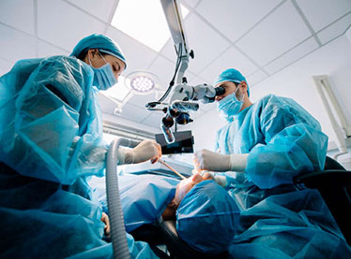
Under local anaesthesia, a flap of gum tissue is elevated to expose the deficient bone. Graft material is placed to fill the defect, a membrane is positioned over the graft to exclude soft tissue ingrowth, and the flap is sutured closed over the site. The entire procedure for a single-site socket preservation takes 30-45 minutes; larger ridge augmentations take 90-120 minutes.
The procedure begins with local anaesthetic infiltration and nerve block as appropriate for the surgical site. A full-thickness mucoperiosteal flap is elevated, meaning the gum tissue and the periosteum (the cellular envelope of the bone) are reflected together, preserving the blood supply to the underlying bone and maximising the healing potential of the surgical site. Decorticating the cortical bone surface with a small round bur or piezo tip activates the regional acceleratory phenomenon (RAP), increasing local blood flow and growth factor availability at the graft site.
Graft material is hydrated with sterile saline or patient blood and packed into the defect. For guided bone regeneration (GBR), a membrane is trimmed and adapted over the graft to prevent fibrous connective tissue, which migrates faster than bone, from colonising the space before osteogenesis can occur. Resorbable collagen membranes (Bio-Gide) integrate and resorb over 12-24 weeks; non-resorbable membranes (titanium-reinforced d-PTFE) maintain space more rigidly but require a second procedure for removal at 6-9 months. The flap is then advanced and sutured with primary tension-free closure, the most critical technical step, as membrane exposure is the leading cause of graft failure.
You should know that primary wound closure, complete coverage of the graft and membrane with sutured gum tissue, is not always achievable without periosteal releasing incisions, which extend the flap and allow tension-free advancement. If your surgeon cannot achieve primary closure, the risk of membrane exposure and graft failure increases substantially. Ask your surgeon how they ensure primary closure at the graft site and whether they routinely use a periosteal release.
At Stunning Dentistry, we perform GBR procedures under optical magnification and confirm dimensional graft thickness intraoperatively with a calibrated probe. All flap designs include a periosteal scoring incision when required for closure, and sutured margins are verified to be tension-free before the patient leaves the surgical suite. Post-operative CBCT is not routine for simple socket preservation but is performed at 6 months post-graft for complex ridge augmentations to confirm bone volume before implant planning resumes.
| Procedural Phase | What Occurs | Duration |
|---|---|---|
| Anaesthesia | Local infiltration + nerve block | 5-10 min |
| Flap elevation | Mucoperiosteal reflection to expose bone | 10-15 min |
| Site preparation | Cortical decortication, defect measurement | 5-10 min |
| Graft placement | Particulate packing or block fixation + membrane | 15-30 min |
| Wound closure | Tension-free suturing with periosteal release as needed | 15-20 min |
| Total (socket preservation) | Single site | ~45 min |
| Total (ridge augmentation) | Moderate-complex | 90-120 min |
Ready to discuss your options?
Pain and Recovery: What to Expect
How much pain is there after a bone graft?
Most patients describe bone grafting recovery as comparable to a tooth extraction, localised soreness, swelling, and bruising for 3-7 days managed with prescribed analgesics. Significant pain beyond day 3-4 is uncommon and warrants clinical assessment. The healing phase before implant placement (3-9 months) is uneventful for most patients; the graft itself is not painful during maturation.
Post-operative discomfort following bone grafting is predictable in intensity and duration. Swelling peaks at 48-72 hours and resolves over 5-7 days; bruising, if present, clears within 10-14 days. Prescribed analgesics, typically ibuprofen 400-600mg every 6-8 hours with paracetamol as required, manage the inflammatory phase in most cases without requiring opioid medication. Ice application during the first 24 hours and elevation of the head at rest reduces oedema formation.
The immediate post-surgical restriction is dietary: soft foods only for 2-3 weeks to avoid mechanical disruption of the suture line and graft site. No tooth brushing directly over the surgical area for 2 weeks; chlorhexidine gluconate rinses replace mechanical cleaning at that site. Sutures are removed at 10-14 days. The patient then enters the graft maturation phase, a period during which there are no clinical appointments and no symptoms; the bone is simply forming at a microscopic level within the scaffold.
You should plan your activity calendar around the first week of recovery rather than the total healing timeline. The 3-9-month maturation period before implant placement is a waiting phase, not a recovery phase. During this time, normal diet, exercise, and daily activity resume fully. The only restriction during maturation is avoidance of removable prostheses (full dentures) that apply pressure over the graft site, as sustained compression can impair bone formation. Your surgeon should provide a vacuum-formed temporary that rests above the graft site rather than loads it.
At Stunning Dentistry, we provide all patients with a written post-operative protocol, a 48-hour post-procedure phone follow-up from our clinical coordinator, and a 2-week suture removal appointment. Patients undergoing complex ridge augmentation are reviewed at 1 week, 1 month, 3 months, and 6 months before CBCT is obtained to confirm graft volume. Remote patients receive CBCT referral instructions for a local imaging centre in the UK, with images transmitted to our surgical team for review before travel is booked for implant placement.
| Recovery Phase | Typical Experience | Required Actions |
|---|---|---|
| Day 1-3 | Swelling, soreness, bruising | Ice, elevation, prescribed analgesics |
| Day 3-14 | Swelling resolves, soreness diminishes | Soft diet, chlorhexidine rinse |
| Day 14 | Suture removal | Clinical review |
| Month 1-3 | No symptoms; graft integrating | Avoid pressure over site; normal activity |
| Month 3-6 | Bone formation active | CBCT at 6 months for complex cases |
| Month 6-9 | Graft maturation | Implant placement appointment booked |
Curious about costs and timelines?
Risk Transparency
What can go wrong with a bone graft?
The most common complication is membrane exposure, partial uncovering of the barrier membrane through the gum tissue, occurring in approximately 15-20% of non-resorbable membrane cases and less frequently with resorbable membranes. Exposed membranes are managed by trimming the exposed portion and prescribing chlorhexidine; graft salvage is often possible. Complete graft failure, infection requiring removal, and nerve proximity complications are less common but occur in complex augmentations.
Membrane exposure is the most clinically significant complication of GBR bone grafting, arising when the sutured tissue margin separates, allowing the underlying membrane to contact the oral environment. The exposed membrane becomes colonised with oral bacteria, which can compromise the underlying graft. With resorbable collagen membranes, early exposure (< 3 weeks) typically requires close monitoring and antimicrobial rinses; late exposure is less consequential as the membrane has already served its space-maintaining function. Non-resorbable membranes (d-PTFE, titanium-reinforced) exposed before 6 months require removal of the exposed portion, which may compromise graft space maintenance and dimensional outcome.
Infection following bone grafting presents as increasing rather than decreasing pain after day 3-4, localised swelling that worsens rather than resolves, and occasionally purulent discharge. True graft infections requiring complete graft removal occur in approximately 3-5% of cases in published series; they are more common in immunocompromised patients, smokers, poorly controlled diabetics, and patients on bisphosphonate therapy. Partial graft salvage following early debridement is possible in many cases where infection is identified and treated promptly.
You should disclose all medications to your surgeon before bone grafting. Bisphosphonates (alendronate, zoledronic acid) taken for osteoporosis or oncological indications alter bone remodelling in ways that significantly affect graft integration and create a risk of medication-related osteonecrosis of the jaw (MRONJ). Systemic steroids, immunosuppressants, and anticoagulants all require specific management protocols around surgical bone procedures. Smoking reduces blood supply to the healing graft bed and doubles the risk of membrane exposure; cessation for at least 2 weeks pre-operatively and throughout the maturation period is strongly recommended.
At Stunning Dentistry, we perform pre-surgical medical history review, medication reconciliation, and risk stratification for every bone grafting patient. High-risk patients receive antibiotic prophylaxis, modified flap designs to maximise primary closure, and more frequent post-operative monitoring. Our surgical team discusses the specific risk profile for each patient's case type before consent is signed, so patients understand not only population-level risks but the factors that modify their individual risk.
| Complication | Frequency | Management | Effect on Outcome |
|---|---|---|---|
| Membrane exposure (resorbable) | 5-10% | Chlorhexidine rinse, monitoring | Often salvageable |
| Membrane exposure (non-resorbable) | 15-20% | Trim exposure, consider early removal | Partial volume loss possible |
| Graft infection | 3-5% | Debridement ± antibiotics ± removal | Variable; early treatment often preserves graft |
| Nerve proximity (mandible) | Site-dependent | Preoperative CBCT nerve mapping | Prevented by planning |
| Sinus membrane perforation (maxilla) | 10-30% in sinus grafts | Collagen plug, resorbable membrane | Manageable intraoperatively |
| Complete graft failure | 2-4% | Repeat augmentation after 3 months | Delays timeline by 3-6 months |
Want a personalised treatment plan?
Clinical Success Determinants
Bone graft success rates in the published surgical literature, defined as adequate volume for planned implant placement without additional augmentation, range from 85-97% for socket preservation and horizontal GBR to 70-85% for complex vertical augmentations. The variability reflects defect size, technique complexity, and patient selection rather than biological unpredictability. When patients are appropriately selected and surgical technique is precise, grafting is a highly reliable preparatory procedure.
| Success Factor | Why It Matters | What to Verify |
|---|---|---|
| Primary wound closure | Exposed membrane leads to bacterial contamination and graft loss | Ask if periosteal release is routinely used |
| Surgeon experience with the specific technique | Vertical GBR and block grafts are technically demanding | Ask how many they perform annually |
| Membrane selection | Space maintenance vs resorption profile must match defect | Confirm membrane type and rationale |
| No smoking during maturation | Smoking impairs angiogenesis critical for graft integration | Cessation for full maturation period |
| Controlled diabetes (HbA1c < 7.5%) | Hyperglycaemia impairs bone healing and increases infection risk | Obtain medical clearance before surgery |
| No bisphosphonate exposure | IV bisphosphonates dramatically increase MRONJ risk | Full medication history required |
| Stable systemic health | ASA class III-IV requires anaesthesia and medical co-management | Medical clearance before surgery |
| Adequate graft volume | Under-packing leads to insufficient bone; over-packing causes membrane tension | Surgeon should measure pre and intra-operatively |
| Patient compliance with soft-diet restriction | Mechanical force disrupts early scaffold before osteoid bridges form | Written protocol and follow-up call |
Questions about this procedure?
Healing Timeline
Socket preservation (small-volume graft at extraction): most cases implant-ready at 3-4 months.
Horizontal GBR (moderate defect): implant placement typically at 5-6 months post-graft.
Lateral sinus lift (large augmentation): staged with implant placement at 6-9 months after the lift.
Vertical ridge augmentation (complex): implant placement at 9-12 months is common.
Timing is assessed by CBCT, not by calendar alone. A graft that appears small can mature rapidly in a young, non-smoking patient; a large augmentation in a poorly controlled diabetic may require extended healing. Implant placement should not proceed until radiographic evidence of bone mineralisation within the grafted volume is confirmed.
| Phase | Timeline | Biological Activity | Clinical Marker |
|---|---|---|---|
| Clot stabilisation | Day 0-7 | Fibrin clot organises; initial scaffold vascularisation | No symptoms; avoid dislodging |
| Early integration | Week 2-6 | Osteoid matrix deposition begins at graft-host interface | Sutures removed; soft diet continues |
| Active bone formation | Month 2-4 | Osteoblasts populate scaffold; mineralisation progresses | No clinical signs; patient asymptomatic |
| Scaffold remodelling | Month 4-6 | Xenograft particles slowly incorporated; bone bridges form | CBCT at 6 months for complex cases |
| Maturation | Month 6-9 | Corticalisation of new bone; adequate density for implant load | Implant planning resumes |
| Late maturation | Month 9-12+ | Required for vertical augmentations and large block grafts | Later CBCT if complex augmentation |
Ready to discuss your options?
When Bone Grafting Is Not Recommended
Who should not have a bone graft?
Bone grafting is contraindicated in patients on IV bisphosphonate therapy (high MRONJ risk), active chemotherapy or radiation to the jaw (impaired wound healing), uncontrolled systemic conditions (HbA1c > 8%, poorly controlled bleeding disorders), or active oral infection at the graft site. Relative contraindications include smoking (cessation required), oral bisphosphonate use (risk stratification required), and thin/compromised soft tissue coverage.
Active malignancy involving the jaw or prior radiation therapy to the head and neck region (> 50 Gy) is a contraindication to elective bone grafting due to the risk of osteoradionecrosis and severely compromised wound healing. Radiation damages the microvascular supply of bone in a way that substantially reduces healing potential for years after the treatment course. In such cases, implant-supported prosthetics may be possible only via zygomatic or pterygoid implants that bypass the irradiated field.
Intravenous bisphosphonate therapy (zoledronic acid, pamidronate) administered for osteoporosis or bone metastases significantly elevates MRONJ risk following any surgical bone procedure. The American Association of Oral and Maxillofacial Surgeons recommends that elective dentoalveolar surgery in patients on IV antiresorptive therapy be approached with extreme caution and multidisciplinary oncological consultation. Oral bisphosphonate users (alendronate, risedronate) represent a lower-risk population, but drug holiday discussions and risk stratification are appropriate for cases requiring extensive grafting.
You should not proceed with bone grafting if there is unresolved periodontal disease, untreated periapical pathology at adjacent teeth, or active infection at the intended graft site. Introducing graft material into a bacterially contaminated field predictably results in failure. All active disease must be treated and resolved, with confirmed healing documented radiographically, before graft surgery is scheduled.
At Stunning Dentistry, we do not proceed with bone grafting until the patient has cleared a structured pre-surgical health review. This includes medical history verification, radiographic confirmation that no active infection exists at the site, medication reconciliation, and, for any patient with systemic health complexity, clearance from the treating physician. Patients who are not suitable candidates at the time of evaluation receive a clear explanation of what must change before surgery can be reconsidered.
| Contraindication | Classification | Clinical Consequence |
|---|---|---|
| IV bisphosphonate therapy | Absolute | MRONJ risk; surgery contraindicated without oncology input |
| Head/neck radiation > 50 Gy | Absolute | Osteoradionecrosis risk; no elective surgery |
| Active oral infection at site | Absolute | Graft failure predictable; resolve first |
| Active malignancy of jaw | Absolute | Surgical wound healing impaired |
| Uncontrolled diabetes (HbA1c > 8%) | Relative | Delayed healing; optimise before surgery |
| Active smoker | Relative | Doubles membrane exposure risk; cessation required |
| Oral bisphosphonate (> 4 years) | Relative | Risk stratification required; possible drug holiday |
| Immunosuppression | Relative | Infection risk elevated; specialist co-management |
Curious about costs and timelines?
Alternative and Adjunct Options
When bone volume is insufficient for standard implants but bone grafting is contraindicated or the patient declines the staged approach, several alternatives exist. Short implants (4-6mm) have shown acceptable survival rates in limited-height bone, published 5-year survival rates of 92-96% in posterior areas, reducing the need for sinus augmentation in many posterior maxillary cases. Narrow-diameter implants expand options in thin ridges but carry higher fracture risk under heavy occlusal loads. These are compromises that accept reduced implant dimensions in exchange for eliminating grafting; they are appropriate for specific anatomical situations, not blanket alternatives.
Zygomatic implants bypass the maxillary sinus and alveolar ridge entirely, anchoring in the zygoma (cheekbone). They are indicated for severely resorbed maxillae where conventional grafting would require multiple staged augmentations and are reserved for specialists trained in the technique. All-on-4 implant design uses angled posterior implants to maximise available anterior-posterior spread without posterior bone grafting, this is the rationale behind the tilted-implant protocol.
You should discuss these alternatives directly in the context of your specific bone defect dimensions, your health profile, and the timeline you are working within. There is no universal answer to whether a short implant, a zygomatic implant, or a staged graft is the right choice, the answer depends on CBCT anatomy, occlusal load requirements, systemic health, and your priorities around surgical complexity and timeline.
At Stunning Dentistry, we present all viable treatment pathways for patients with bone-deficient ridges, including the clinical rationale for each option, the data on outcomes at equivalent follow-up, and the specific limitations or unknowns for the patient's anatomy. Patients are not guided toward grafting unless the clinical evidence supports it as the best path for their specific case.
Want a personalised treatment plan?
Cost Logic: GBP Comparison
Materials note: Stunning Dentistry uses the same graft materials available in British oral surgery practices, Geistlich Bio-Oss, Geistlich Bio-Gide, MinerOss allograft, Osteon III, sourced from the same global suppliers. The cost differential reflects surgical facility overhead, anaesthesia billing structures, and specialist fee schedules, not material or technical standard.
Wait time note: Specialist oral surgeon consults in the UK carry average wait times of 3-8 weeks in major cities, with surgical bookings often 4-12 weeks beyond that. Patients travelling to Stunning Dentistry receive a CBCT-informed treatment plan within 12 hours of arrival and surgery within the first clinical week.
Total trip cost: For patients whose bone grafting is part of a full-mouth implant treatment plan, the combined treatment-and-travel cost in India typically remains significantly lower than the isolated surgical cost at a British oral surgery practice, even accounting for flights and accommodation.
| Procedure | Stunning Dentistry (India), GBP | British Oral Surgeon, GBP | Difference |
|---|---|---|---|
| Socket preservation (1 site) | £500 - £750 | £900 - £1,500 | ~£400 - £750 saved |
| Horizontal ridge augmentation (1 site) | £750 - £1,200 | £1,500 - £3,500 | ~£750 - £1,800 saved |
| Vertical ridge augmentation | £1,200 - £2,100 | £3,500 - £4,800+ | ~£1,800 - £3,500 saved |
| Lateral sinus lift (1 side) | from £210 | £1,800 - £3,600 | ~£900 - £2,100 saved |
| Transcrestal sinus lift (1 site) | from £210 | £750 - £1,500 | ~£360 - £900 saved |
| Full arch bone grafting (complex) | from £210 | £8,700 - £18,000+ | ~£6,000 - £12,000 saved |
Questions about this procedure?

Treatment Comparison Matrix
| Factor | GBR (Particulate + Membrane) | Block Autograft | Short Implants | Zygomatic Implants |
|---|---|---|---|---|
| Indication | Horizontal defects up to 4-5mm | Vertical defects; large horizontal | Sub-optimal bone height; posterior | Severely resorbed maxilla |
| Second surgical site | No (with xenograft) | Yes (mandible/iliac) | No | No |
| Staging required | Yes: 3-6 months | Yes: 6-12 months | No (immediate placement possible) | No (single surgery) |
| Complication profile | Membrane exposure | Donor site morbidity; block resorption | Higher fracture risk (narrow implants) | Technical demands; sinus adjacency |
| Long-term bone volume | Stable with xenograft | Variable; some resorption | Not applicable (no graft) | Not applicable |
| Cost (GBP, SD) | £750-£1,500 | £1,500-£2,600 | Lower (no graft fee) | Higher (complex surgery) |
| Best for | Most GBR candidates | Large vertical deficiencies | Low-risk posterior cases | Failed graft, severe resorption |
Ready to discuss your options?
Post-Graft Biological Reality
The bone that forms within a graft site is not identical to the bone that was originally present. New bone forms as the graft scaffold is populated by osteoblasts migrating from the adjacent host bone margins and from marrow-derived precursor cells mobilised by the surgical stimulus. Over time, the new bone remodels into lamellar architecture, the organised, load-bearing structure of mature cortical bone, but this process takes 12-18 months in larger augmentations, not just the 6-9 months before implant placement.
Xenograft particles (Bio-Oss) do not fully resorb within the clinical timeframe. They remain as slowly resorbing mineral particles within the new bone matrix, contributing to long-term ridge volume maintenance. Histological studies have confirmed osseointegration of Bio-Oss particles within the newly formed bone, they are incorporated rather than excluded. This is a feature, not a limitation: xenograft-supported grafted ridges show lower long-term bone volume loss than sites grafted with fully resorbable materials.
You should understand that the implant placed in grafted bone is supported by the new bone, not by the graft material itself. Implant osseointegration depends on direct bone-to-implant contact in the mineralised regions of the grafted volume. This is why CBCT confirmation of graft maturation before implant placement matters: an implant placed too early, before the graft is adequately mineralised, will not achieve the primary stability threshold required for predictable osseointegration.
At Stunning Dentistry, we do not place implants in grafted sites based on elapsed time alone. CBCT radiodensity measurement (Hounsfield units) within the grafted volume guides timing. Sites that show adequate mineralisation at 4 months are implant-staged at 5 months; sites with slower maturation are held at 8-9 months. This protects implant success rates in the grafted bone rather than applying a blanket calendar protocol.
Curious about costs and timelines?
Common Mistakes That Compromise Grafts
What causes bone graft failure?
The majority of bone graft failures are attributable to preventable technical or patient-compliance factors: inadequate primary wound closure, pressure from removable dentures over the graft site, smoking during the maturation period, placing implants before radiographic confirmation of graft maturation, and inadequate membrane selection for the defect geometry.
The most consequential intraoperative mistake is failure to achieve tension-free primary closure. Surgeons who underestimate the importance of periosteal release accept wound margins under tension; within 72-96 hours, the sutures pull through under normal swelling pressure, exposing the membrane. Membrane exposure is not always a full graft failure, but it consistently reduces the volume of bone formed. The primary closure step is where surgical skill most directly impacts graft outcome.
Placing a removable denture or retainer over the graft site in the early maturation phase applies compressive force to the forming bone scaffold. Bone formation requires an undisturbed mechanical environment; sustained pressure inhibits osteoblast activity and collapses the membrane's space-maintaining function. Patients who cannot be without a denture need a vacuum-formed or implant-supported temporary specifically designed to float above the graft site rather than rest on it.
You should not expect that a failed or partial graft is necessarily irreversible. Secondary augmentation at a previously grafted site, performed after allowing the failed graft to stabilise and any infection to resolve, is routinely performed. Success rates for secondary grafting are lower than for primary grafting but remain clinically significant. A partial graft that achieved some volume reduction but not full augmentation can sometimes be combined with a shorter or narrower implant design to avoid complete re-augmentation.
At Stunning Dentistry, we review all patients for removable prosthesis compatibility before graft surgery and replace any appliance that would load the graft site. Patients who require an immediate temporary for aesthetic reasons receive a custom vacuum-formed appliance that is relieved over the surgical site before the grafting appointment. Adherence to soft-diet restriction and denture non-use is confirmed at each post-operative visit.
Want a personalised treatment plan?
Myth Deconstruction
Myth: "If the graft fails, I can never get implants."
A failed graft does not eliminate implant options. Secondary augmentation can be performed after allowing the site to stabilise. Zygomatic implants, pterygoid implants, or short-implant designs may provide viable alternatives depending on the residual anatomy. Failed bone grafting is a setback in timeline and cost, not an irreversible outcome in most cases.
Myth: "Bone grafting always requires bone from my hip."
The large majority of dental bone grafting procedures use xenograft (bovine bone mineral), allograft (processed human donor bone), or synthetic materials, none of which require a second surgical site. Autogenous bone from the hip (iliac crest) is reserved for the most complex large-volume vertical augmentations and represents a small fraction of total grafting volume in implant dentistry.
Myth: "The newer the graft material, the better the outcome."
Geistlich Bio-Oss, introduced commercially in the 1990s, remains the most documented xenograft material in implant dentistry with over 30 years of clinical follow-up data. Newer synthetic and growth-factor-enhanced materials may offer advantages in specific situations but lack the long-term outcome literature of established materials. Clinical evidence depth matters as much as material novelty.
Myth: "Bone grafting is only needed for older patients."
Bone grafting need is determined by ridge dimensions and implant planning requirements, not age. A 28-year-old who lost a tooth from trauma and waited two years before seeking treatment may have a more significantly resorbed ridge than a 60-year-old who received socket preservation at the time of extraction. Age affects healing rate; it does not determine whether grafting is needed.
Questions about this procedure?

People Also Ask
How long does a bone graft last before implant placement?
Bone grafts are not temporary; once integrated, the new bone is permanent. The waiting period before implant placement, 3-9 months depending on graft type and defect size, is not the lifespan of the graft. It is the maturation period during which graft material is replaced by or integrated with new mineralised bone. Once an implant is placed and osseointegrated, the grafted bone behaves like native bone under functional loading.
Can I eat normally after a bone graft?
Soft diet is required for 2-3 weeks post-surgery to protect the wound margins and graft site from mechanical disruption. After suture removal at 2 weeks, most foods can be reintroduced gradually except for hard, crunchy, or chewy items directly over the surgical site. Normal unrestricted eating resumes when the surgeon confirms adequate healing, typically by 4-6 weeks post-graft.
Is bone grafting painful?
Immediate post-surgical soreness, swelling, and bruising are expected for 3-7 days and are managed with prescribed anti-inflammatory analgesics. The bone maturation phase over the following months is asymptomatic, patients describe no ongoing discomfort during the healing period. Significant or worsening pain after day 4 is not expected and should be reported for clinical review.
Do I need a bone graft for every dental implant?
No. Bone grafting is indicated only when CBCT analysis confirms insufficient bone volume for the planned implant. Patients with adequate bone can proceed directly to implant placement. The proportion of implant patients who require some form of grafting is approximately 35-40% in published surgical series, reflecting the prevalence of post-extraction bone loss.
What is the difference between a sinus lift and a bone graft?
A sinus lift is a specific type of bone grafting procedure for the posterior upper jaw, where the maxillary sinus has expanded (pneumatised) into the space previously occupied by teeth and their supporting bone. The sinus membrane is elevated and graft material is placed beneath it to restore sub-sinus bone height. All sinus lifts are bone graft procedures; not all bone graft procedures are sinus lifts.
Will my body reject the bone graft material?
Processed xenograft and allograft materials have had the organic cellular components (antigens) removed, eliminating immunological rejection risk. These materials are biocompatible mineral scaffolds, not transplanted tissue. True immunological rejection does not occur with commercially processed graft materials. Infection and mechanical failure are the relevant risks, not rejection.
Ready to discuss your options?
Ask Your Doctor
- What type of bone graft material will be used for my case, and why is that specific material indicated for my defect?
- Will you use a resorbable or non-resorbable membrane, and what are the implications if it becomes exposed?
- How do you ensure primary wound closure in my case, will you need a periosteal releasing incision?
- How many GBR or ridge augmentation procedures do you perform annually, and what is your graft success rate?
- What does graft success or partial success mean in my case, and what are the options if volume is insufficient after healing?
- When and how will you confirm that the graft has matured adequately before planning implant placement?
- Will I need to avoid wearing my denture over the graft site, and if so, what will I wear in the interim?
- What medications do I need to stop before surgery, and for how long?
Curious about costs and timelines?
Want a personalised treatment plan?
For British Patients
Pre-Travel Checklist for British Patients Considering Bone Grafting in India
Dental Angel Handover: Stunning Dentistry provides a structured clinical handover document for your local British dentist covering graft type used, membrane type and removal requirements (if non-resorbable), post-operative protocol, and CBCT imaging schedule. This document is in English and formatted for acceptance by British dental practices.
| Step | Action | When |
|---|---|---|
| 1 | Obtain CBCT scan at British imaging centre (e.g., OmegaDent, Radiologix) | Before booking travel |
| 2 | Send DICOM files to Stunning Dentistry for remote treatment planning | 4-6 weeks before travel |
| 3 | Receive written treatment plan with graft type, timeline, and GBP costs | Pre-travel |
| 4 | Obtain medical clearance from GP if on bisphosphonates, anticoagulants, or immunosuppressants | Pre-travel |
| 5 | Confirm soft foods are accessible at accommodation for 2 weeks post-surgery | Pre-travel |
| 6 | Arrange local dentist in the UK for suture removal (if returning before 14-day post-op) | Pre-travel |
| 7 | Arrange local imaging centre for 6-month post-graft CBCT (transmitted to SD for review) | Pre-travel |
| 8 | Plan return visit for implant placement after CBCT confirms graft maturation | At 6-9 months |
Questions about this procedure?
Clinical References
1. Araújo MG, Lindhe J. "Dimensional ridge alterations following tooth extraction." *Journal of Clinical Periodontology* 2005;32(2):212-218.
*Medically Reviewed, Stunning Dentistry Oral Surgery Department. Protocols consistent with ITI Consensus Statements on bone augmentation for implant dentistry. Content reviewed May 2026.*
Ready to discuss your options?
Ready to Start?
Every case is planned by a named lead clinician and backed by a written Lifetime Warranty. Share your scans or a photo for a no-obligation clinical assessment.
Curious about costs and timelines?
Specialist-only treatment planning
- Remote file review before travel
- Evidence-led treatment checkpoints
No waiting list for eligible cases
- Remote file review before travel
- Evidence-led treatment checkpoints
Trip coordinated with care timeline
- Remote file review before travel
- Evidence-led treatment checkpoints
Our Partners











Why Us
Frequently Asked Questions
Can bone grafting and implant placement happen at the same time?
Simultaneous grafting and implant placement (immediate placement with simultaneous GBR) is possible when the implant achieves adequate primary stability and the bone defect is a contained buccal gap defect rather than a severe ridge deficiency. This approach shortens overall treatment time but requires careful case selection. Where primary stability cannot be confirmed or the defect is too large, staged grafting provides more reliable outcomes.
What happens to a bone graft if I don't end up getting the implant?
The grafted bone remains as permanent augmented ridge regardless of whether an implant is placed. It does not resorb specifically because an implant was not placed. However, over years without functional stimulation (from an implant or a tooth), any augmented ridge will undergo some degree of resorption, gradually reverting toward its pre-graft dimensions. This is why grafting should be followed by implant placement within a clinically appropriate window rather than deferred indefinitely.
Is bone grafting covered by British insurance?
Coverage varies significantly. Some extended health benefit plans cover surgical procedures including bone grafting at a percentage of the fee guide; others specifically exclude implant-preparatory procedures. The NHS or private dental cover does not cover elective dental procedures. Check your plan's specific language around oral surgery, bone augmentation, and implant preparatory procedures before assuming coverage.
How do I know if my graft has failed?
Early signs include significant pain or swelling after day 4-5, membrane exposure that does not respond to management, or purulent discharge from the surgical site. Late graft failure may present as insufficient bone on the CBCT obtained at 6 months, the grafted volume is present but not mineralised. A CBCT obtained by a qualified radiologist before implant placement is the definitive method of assessing graft maturity and volume adequacy.
See your new smile instantly!
This tool will help you understand potential structural and aesthetic changes before finalizing treatment decisions.


